

64
4 Synthesis of Two-Dimensional (2D) Nanomaterials
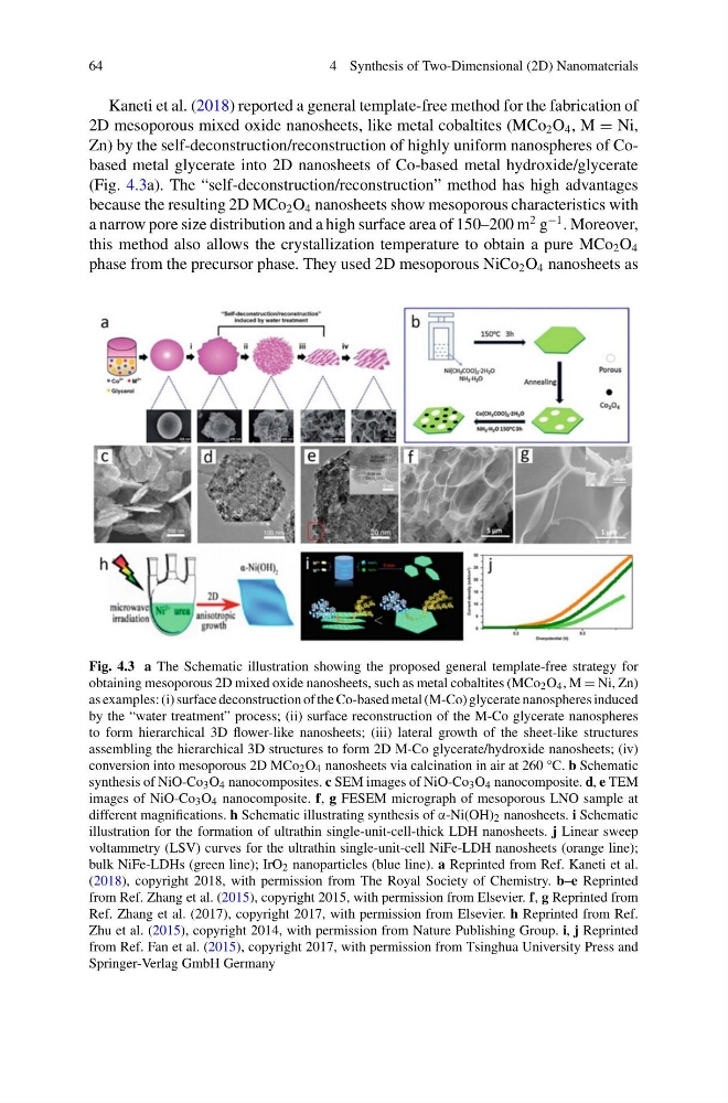
Kaneti et al. (2018) reported a general template-free method for the fabrication of
2D mesoporous mixed oxide nanosheets, like metal cobaltites (MCo 2 O 4 , M = Ni,
Zn) by the self-deconstruction/reconstruction of highly uniform nanospheres of Cobased metal glycerate into 2D nanosheets of Co-based metal hydroxide/glycerate
(Fig. 4.3a). The “self-deconstruction/reconstruction” method has high advantages
because the resulting 2D MCo 2 O 4 nanosheets show mesoporous characteristics with
a narrow pore size distribution and a high surface area of 150–200 m
2 g
−1 . Moreover,
this method also allows the crystallization temperature to obtain a pure MCo 2 O 4
phase from the precursor phase. They used 2D mesoporous NiCo 2 O 4 nanosheets as
Fig. 4.3 a The Schematic illustration showing the proposed general template-free strategy for
obtaining mesoporous 2D mixed oxide nanosheets, such as metal cobaltites (MCo 2 O 4 , M = Ni, Zn)
as examples: (i) surface deconstruction of the Co-based metal (M-Co) glycerate nanospheres induced
by the “water treatment” process; (ii) surface reconstruction of the M-Co glycerate nanospheres
to form hierarchical 3D flower-like nanosheets; (iii) lateral growth of the sheet-like structures
assembling the hierarchical 3D structures to form 2D M-Co glycerate/hydroxide nanosheets; (iv)
conversion into mesoporous 2D MCo 2 O 4 nanosheets via calcination in air at 260 °C. b Schematic
synthesis of NiO-Co 3 O 4 nanocomposites. c SEM images of NiO-Co 3 O 4 nanocomposite. d, e TEM
images of NiO-Co 3 O 4 nanocomposite. f, g FESEM micrograph of mesoporous LNO sample at
different magnifications. h Schematic illustrating synthesis of α-Ni(OH) 2 nanosheets. i Schematic
illustration for the formation of ultrathin single-unit-cell-thick LDH nanosheets. j Linear sweep
voltammetry (LSV) curves for the ultrathin single-unit-cell NiFe-LDH nanosheets (orange line);
bulk NiFe-LDHs (green line); IrO 2 nanoparticles (blue line). a Reprinted from Ref. Kaneti et al.
(2018), copyright 2018, with permission from The Royal Society of Chemistry. b–e Reprinted
from Ref. Zhang et al. (2015), copyright 2015, with permission from Elsevier. f, g Reprinted from
Ref. Zhang et al. (2017), copyright 2017, with permission from Elsevier. h Reprinted from Ref.
Zhu et al. (2015), copyright 2014, with permission from Nature Publishing Group. i, j Reprinted
from Ref. Fan et al. (2015), copyright 2017, with permission from Tsinghua University Press and
Springer-Verlag GmbH Germany
4 Synthesis of Two-Dimensional (2D) Nanomaterials
Kaneti et al. (2018) reported a general template-free method for the fabrication of
2D mesoporous mixed oxide nanosheets, like metal cobaltites (MCo 2 O 4 , M = Ni,
Zn) by the self-deconstruction/reconstruction of highly uniform nanospheres of Cobased metal glycerate into 2D nanosheets of Co-based metal hydroxide/glycerate
(Fig. 4.3a). The “self-deconstruction/reconstruction” method has high advantages
because the resulting 2D MCo 2 O 4 nanosheets show mesoporous characteristics with
a narrow pore size distribution and a high surface area of 150–200 m
2 g
−1 . Moreover,
this method also allows the crystallization temperature to obtain a pure MCo 2 O 4
phase from the precursor phase. They used 2D mesoporous NiCo 2 O 4 nanosheets as
Fig. 4.3 a The Schematic illustration showing the proposed general template-free strategy for
obtaining mesoporous 2D mixed oxide nanosheets, such as metal cobaltites (MCo 2 O 4 , M = Ni, Zn)
as examples: (i) surface deconstruction of the Co-based metal (M-Co) glycerate nanospheres induced
by the “water treatment” process; (ii) surface reconstruction of the M-Co glycerate nanospheres
to form hierarchical 3D flower-like nanosheets; (iii) lateral growth of the sheet-like structures
assembling the hierarchical 3D structures to form 2D M-Co glycerate/hydroxide nanosheets; (iv)
conversion into mesoporous 2D MCo 2 O 4 nanosheets via calcination in air at 260 °C. b Schematic
synthesis of NiO-Co 3 O 4 nanocomposites. c SEM images of NiO-Co 3 O 4 nanocomposite. d, e TEM
images of NiO-Co 3 O 4 nanocomposite. f, g FESEM micrograph of mesoporous LNO sample at
different magnifications. h Schematic illustrating synthesis of α-Ni(OH) 2 nanosheets. i Schematic
illustration for the formation of ultrathin single-unit-cell-thick LDH nanosheets. j Linear sweep
voltammetry (LSV) curves for the ultrathin single-unit-cell NiFe-LDH nanosheets (orange line);
bulk NiFe-LDHs (green line); IrO 2 nanoparticles (blue line). a Reprinted from Ref. Kaneti et al.
(2018), copyright 2018, with permission from The Royal Society of Chemistry. b–e Reprinted
from Ref. Zhang et al. (2015), copyright 2015, with permission from Elsevier. f, g Reprinted from
Ref. Zhang et al. (2017), copyright 2017, with permission from Elsevier. h Reprinted from Ref.
Zhu et al. (2015), copyright 2014, with permission from Nature Publishing Group. i, j Reprinted
from Ref. Fan et al. (2015), copyright 2017, with permission from Tsinghua University Press and
Springer-Verlag GmbH Germany