

instead of keeping the holding needle static at the bottom of
the silique, it is recommended to place it close to the incision
point. In that way, after dissecting a short stretch with the
dissecting needle, the holding needle should be moved downward to hold the silique for the next dissection stretch. This
should be repeated until dissection is finished.
5. Siliques tend to ascent to the surface. It is important to ensure
that at least the bases of siliques are immersed in fixing solution.
After a few hours, the penetrated fixing solution will remove all
air inside siliques and they will sink. This can be accelerated by
shaking siliques 2–3 times downward inside tubes.
6. Always harvest and hold the siliques by their pedicels; otherwise tweezers could damage the ovary cavity and the ovules/
seeds inside.
7. Pedicels longer than 5 mm could prevent visualization of both
silique compartments after clearing. In that case, we suggest to
cut down the pedicel (do not remove it completely) inside the
same Petri dish with two high-precision tweezers.
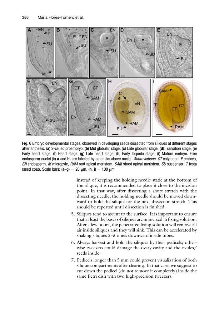
Fig. 6 Embryo developmental stages, observed in developing seeds dissected from siliques at different stages
after anthesis. (a) 2-celled proembryo. (b) Mid globular stage. (c) Late globular stage. (d) Transition stage. (e)
Early heart stage. (f) Heart stage. (g) Late heart stage. (h) Early torpedo stage. (i) Mature embryo. Free
endosperm nuclei (in a and b) are labeled by asterisks above nuclei. Abbreviations: CT cotyledon, E embryo,
EN endosperm, M micropyle, RAM root apical meristem, SAM shoot apical meristem, SU suspensor, T testa
(seed coat). Scale bars: (a–g) ¼ 20 μm, (h, i) ¼ 100 μm
386
Marı ´a Flores-Tornero et al.
the silique, it is recommended to place it close to the incision
point. In that way, after dissecting a short stretch with the
dissecting needle, the holding needle should be moved downward to hold the silique for the next dissection stretch. This
should be repeated until dissection is finished.
5. Siliques tend to ascent to the surface. It is important to ensure
that at least the bases of siliques are immersed in fixing solution.
After a few hours, the penetrated fixing solution will remove all
air inside siliques and they will sink. This can be accelerated by
shaking siliques 2–3 times downward inside tubes.
6. Always harvest and hold the siliques by their pedicels; otherwise tweezers could damage the ovary cavity and the ovules/
seeds inside.
7. Pedicels longer than 5 mm could prevent visualization of both
silique compartments after clearing. In that case, we suggest to
cut down the pedicel (do not remove it completely) inside the
same Petri dish with two high-precision tweezers.
Fig. 6 Embryo developmental stages, observed in developing seeds dissected from siliques at different stages
after anthesis. (a) 2-celled proembryo. (b) Mid globular stage. (c) Late globular stage. (d) Transition stage. (e)
Early heart stage. (f) Heart stage. (g) Late heart stage. (h) Early torpedo stage. (i) Mature embryo. Free
endosperm nuclei (in a and b) are labeled by asterisks above nuclei. Abbreviations: CT cotyledon, E embryo,
EN endosperm, M micropyle, RAM root apical meristem, SAM shoot apical meristem, SU suspensor, T testa
(seed coat). Scale bars: (a–g) ¼ 20 μm, (h, i) ¼ 100 μm
386
Marı ´a Flores-Tornero et al.