

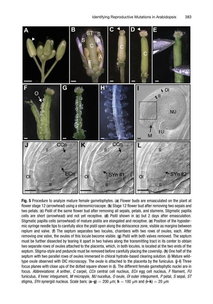
Fig. 5 Procedure to analyze mature female gametophytes. (a) Flower buds are emasculated on the plant at
flower stage 12 (arrowhead) using a stereomicroscope. (b) Stage 12 flower bud after removing two sepals and
two petals. (c) Pistil of the same flower bud after removing all sepals, petals, and stamens. Stigmatic papilla
cells are short (arrowhead) and not yet receptive. (d) Pistil shown in (c) but 2 days after emasculation.
Stigmatic papilla cells (arrowhead) of mature pistils are elongated and receptive. (e) Position of the hypodermic syringe needle tips to carefully slice the pistil open along the dehiscence zone, visible as margins between
replum and valve. (f) The septum separates two locules, chambers with two rows of ovules, each. After
removing one valve, the ovules of this locule become visible. (g) Pistil with both valves removed. The septum
must be further dissected by tearing it apart in two halves along the transmitting tract in its center to obtain
two separate rows of ovules attached to the placenta, which, in both locules, is located at the two ends of the
septum. Stigma-style and peduncle must be removed before carefully placing the coverslip. (h) One half of the
septum with two parallel rows of ovules immersed in chloral hydrate-based clearing solution. (i) Mature wildtype ovule observed with DIC microscopy. The ovule is attached to the placenta by the funiculus. (j–l) Three
focus planes with close ups of the dotted square shown in (i). The different female gametophytic nuclei are in
focus. Abbreviations: A anther, C carpel, CCn central cell nucleus, ECn egg cell nucleus, F filament, FU
funiculus, II Inner integument, M micropyle, NU nucellus, O ovule, OI outer integument, P petal, S sepal, ST
stigma, SYn synergid nucleus. Scale bars: (a–g) ¼ 200 μm; h ¼ 100 μm and (i–k) ¼ 20 μm
Identifying Reproductive Mutations in Arabidopsis
383
flower stage 12 (arrowhead) using a stereomicroscope. (b) Stage 12 flower bud after removing two sepals and
two petals. (c) Pistil of the same flower bud after removing all sepals, petals, and stamens. Stigmatic papilla
cells are short (arrowhead) and not yet receptive. (d) Pistil shown in (c) but 2 days after emasculation.
Stigmatic papilla cells (arrowhead) of mature pistils are elongated and receptive. (e) Position of the hypodermic syringe needle tips to carefully slice the pistil open along the dehiscence zone, visible as margins between
replum and valve. (f) The septum separates two locules, chambers with two rows of ovules, each. After
removing one valve, the ovules of this locule become visible. (g) Pistil with both valves removed. The septum
must be further dissected by tearing it apart in two halves along the transmitting tract in its center to obtain
two separate rows of ovules attached to the placenta, which, in both locules, is located at the two ends of the
septum. Stigma-style and peduncle must be removed before carefully placing the coverslip. (h) One half of the
septum with two parallel rows of ovules immersed in chloral hydrate-based clearing solution. (i) Mature wildtype ovule observed with DIC microscopy. The ovule is attached to the placenta by the funiculus. (j–l) Three
focus planes with close ups of the dotted square shown in (i). The different female gametophytic nuclei are in
focus. Abbreviations: A anther, C carpel, CCn central cell nucleus, ECn egg cell nucleus, F filament, FU
funiculus, II Inner integument, M micropyle, NU nucellus, O ovule, OI outer integument, P petal, S sepal, ST
stigma, SYn synergid nucleus. Scale bars: (a–g) ¼ 200 μm; h ¼ 100 μm and (i–k) ¼ 20 μm
Identifying Reproductive Mutations in Arabidopsis
383