

3. Prepare a microscope glass slide under a stereomicroscope.
4. Take a freshly opened flower (see also Fig. 1b) using tweezers.
5. While holding the flower under the stereomicroscope, dab it
onto the glass slide so you can check how pollen grains are
released onto the surface (see Note 9).
6. Immediately add 7 μL of FDA-BK solution.
7. Place a coverslip on top and incubate in a humid chamber for
5 min in the dark.
8. Observe pollen using a fluorescence microscope with GFP
settings.
9. Compare fluorescence of pollen grains that are viable compared
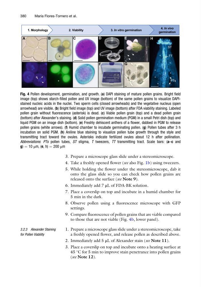
to those that are not viable (Fig. 4b, lower panel).
3.2.3 Alexander Staining
for Pollen Viability
1. Prepare a microscope glass slide under a stereomicroscope, take
a freshly opened flower, and release pollen as described above.
2. Immediately add 5 μL of Alexander stain (see Note 11).
3. Place a coverslip on top and incubate onto a heating surface at
45
C for 5 min to improve stain penetrance into pollen grains
(see Note 12).
Fig. 4 Pollen development, germination, and growth. (a) DAPI staining of mature pollen grains. Bright field
image (top) shows starch-filled pollen and UV image (bottom) of the same pollen grains to visualize DAPIstained nucleic acids in the nuclei. Two sperm cells (closed arrowheads) and the vegetative nucleus (open
arrowhead) are visible. (b) Bright field image (top) and UV image (bottom) after FDA viability staining. Labeled
pollen grain without fluorescence (asterisk) is dead. (c) Viable pollen grain (top) and a dead pollen grain
(bottom) after Alexander’s staining. (d) Solid pollen germination medium (PGM) in a small Petri dish (top) and
liquid PGM on an image dish (bottom). (e) Freshly dehiscent anthers of a flower, dabbed in PGM to release
pollen grains (white arrows). (f) Humid chamber to incubate germinating pollen. (g) Pollen tubes after 3 h
incubation on solid PGM. (h) Aniline blue staining to visualize pollen tube growth through the style and
transmitting tract toward the ovules. Asterisks indicate fertilized ovules about 12 h after pollination.
Abbreviations: PTs pollen tubes, ST stigma, T tweezers, TT transmitting tract. Scale bars: (a–c and
g) ¼ 10 μm, (e, h) ¼ 200 μm
380
Marı ´a Flores-Tornero et al.
4. Take a freshly opened flower (see also Fig. 1b) using tweezers.
5. While holding the flower under the stereomicroscope, dab it
onto the glass slide so you can check how pollen grains are
released onto the surface (see Note 9).
6. Immediately add 7 μL of FDA-BK solution.
7. Place a coverslip on top and incubate in a humid chamber for
5 min in the dark.
8. Observe pollen using a fluorescence microscope with GFP
settings.
9. Compare fluorescence of pollen grains that are viable compared
to those that are not viable (Fig. 4b, lower panel).
3.2.3 Alexander Staining
for Pollen Viability
1. Prepare a microscope glass slide under a stereomicroscope, take
a freshly opened flower, and release pollen as described above.
2. Immediately add 5 μL of Alexander stain (see Note 11).
3. Place a coverslip on top and incubate onto a heating surface at
45
C for 5 min to improve stain penetrance into pollen grains
(see Note 12).
Fig. 4 Pollen development, germination, and growth. (a) DAPI staining of mature pollen grains. Bright field
image (top) shows starch-filled pollen and UV image (bottom) of the same pollen grains to visualize DAPIstained nucleic acids in the nuclei. Two sperm cells (closed arrowheads) and the vegetative nucleus (open
arrowhead) are visible. (b) Bright field image (top) and UV image (bottom) after FDA viability staining. Labeled
pollen grain without fluorescence (asterisk) is dead. (c) Viable pollen grain (top) and a dead pollen grain
(bottom) after Alexander’s staining. (d) Solid pollen germination medium (PGM) in a small Petri dish (top) and
liquid PGM on an image dish (bottom). (e) Freshly dehiscent anthers of a flower, dabbed in PGM to release
pollen grains (white arrows). (f) Humid chamber to incubate germinating pollen. (g) Pollen tubes after 3 h
incubation on solid PGM. (h) Aniline blue staining to visualize pollen tube growth through the style and
transmitting tract toward the ovules. Asterisks indicate fertilized ovules about 12 h after pollination.
Abbreviations: PTs pollen tubes, ST stigma, T tweezers, TT transmitting tract. Scale bars: (a–c and
g) ¼ 10 μm, (e, h) ¼ 200 μm
380
Marı ´a Flores-Tornero et al.