

AFM was initially dedicated to nanoscale imaging, but bigger
indenters were also adapted for AFM, enabling the scan of larger
surfaces at cellular and tissue scales [20, 21]. Currently, AFM
operates with a variety of cantilevers characterized by different
stiffness and tip size and/or shape. The cantilever should be accurate to reflect material properties. The measurement of live and
Detector
Laser
Cantilever
Tip
Glass
slide
Resin embedded
section
A
B
C
D
No contact
(n c)
Contact (c)
No deflection
(n d)
40x / 0.65
Objective
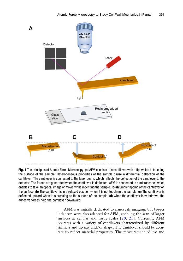
Fig. 1 The principles of Atomic Force Microscopy. (a) AFM consists of a cantilever with a tip, which is touching
the surface of the sample. Heterogeneous properties of the sample cause a differential deflection of the
cantilever. The cantilever is connected to the laser beam, which reflects the deflection of the cantilever to the
detector. The forces are generated when the cantilever is deflected. AFM is connected to a microscope, which
enables to take an optical image or movie while indenting the sample. (b–d) Single tapping of the cantilever on
the surface. (b) The cantilever is in a relaxed position when it is not touching the sample. (c) The cantilever is
deflected upward when it is pressing on the surface of the sample. (d) When the cantilever is withdrawn, the
adhesive forces hold the cantilever downward
Atomic Force Microscopy to Study Cell Wall Mechanics in Plants
351
indenters were also adapted for AFM, enabling the scan of larger
surfaces at cellular and tissue scales [20, 21]. Currently, AFM
operates with a variety of cantilevers characterized by different
stiffness and tip size and/or shape. The cantilever should be accurate to reflect material properties. The measurement of live and
Detector
Laser
Cantilever
Tip
Glass
slide
Resin embedded
section
A
B
C
D
No contact
(n c)
Contact (c)
No deflection
(n d)
40x / 0.65
Objective
Fig. 1 The principles of Atomic Force Microscopy. (a) AFM consists of a cantilever with a tip, which is touching
the surface of the sample. Heterogeneous properties of the sample cause a differential deflection of the
cantilever. The cantilever is connected to the laser beam, which reflects the deflection of the cantilever to the
detector. The forces are generated when the cantilever is deflected. AFM is connected to a microscope, which
enables to take an optical image or movie while indenting the sample. (b–d) Single tapping of the cantilever on
the surface. (b) The cantilever is in a relaxed position when it is not touching the sample. (c) The cantilever is
deflected upward when it is pressing on the surface of the sample. (d) When the cantilever is withdrawn, the
adhesive forces hold the cantilever downward
Atomic Force Microscopy to Study Cell Wall Mechanics in Plants
351