

4. Stain sections by floating grids on a drop of freshly prepared
Reynold’s lead citrate for 2–5 min. and rinse with distilled
water for 30 s (see Note 9).
5. Image stained sections in a regular transmission electron
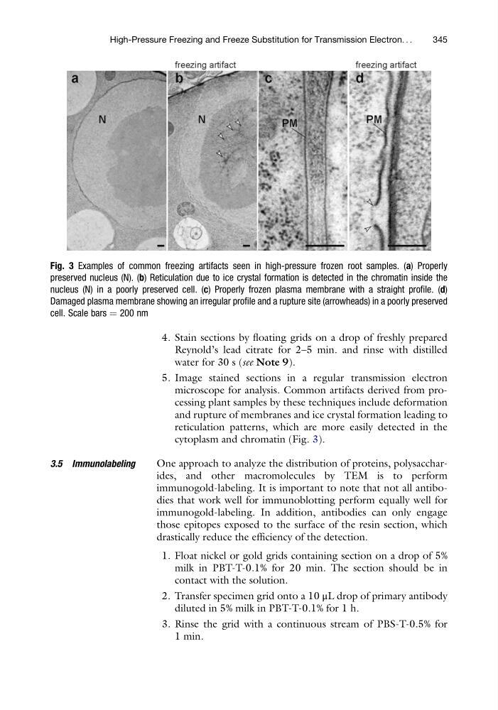
microscope for analysis. Common artifacts derived from processing plant samples by these techniques include deformation
and rupture of membranes and ice crystal formation leading to
reticulation patterns, which are more easily detected in the
cytoplasm and chromatin (Fig. 3).
3.5 Immunolabeling
One approach to analyze the distribution of proteins, polysaccharides, and other macromolecules by TEM is to perform
immunogold-labeling. It is important to note that not all antibodies that work well for immunoblotting perform equally well for
immunogold-labeling. In addition, antibodies can only engage
those epitopes exposed to the surface of the resin section, which
drastically reduce the efficiency of the detection.
1. Float nickel or gold grids containing section on a drop of 5%
milk in PBT-T-0.1% for 20 min. The section should be in
contact with the solution.
2. Transfer specimen grid onto a 10 μL drop of primary antibody
diluted in 5% milk in PBT-T-0.1% for 1 h.
3. Rinse the grid with a continuous stream of PBS-T-0.5% for
1 min.
Fig. 3 Examples of common freezing artifacts seen in high-pressure frozen root samples. (a) Properly
preserved nucleus (N). (b) Reticulation due to ice crystal formation is detected in the chromatin inside the
nucleus (N) in a poorly preserved cell. (c) Properly frozen plasma membrane with a straight profile. (d)
Damaged plasma membrane showing an irregular profile and a rupture site (arrowheads) in a poorly preserved
cell. Scale bars ¼ 200 nm
High-Pressure Freezing and Freeze Substitution for Transmission Electron. . .
345
Reynold’s lead citrate for 2–5 min. and rinse with distilled
water for 30 s (see Note 9).
5. Image stained sections in a regular transmission electron
microscope for analysis. Common artifacts derived from processing plant samples by these techniques include deformation
and rupture of membranes and ice crystal formation leading to
reticulation patterns, which are more easily detected in the
cytoplasm and chromatin (Fig. 3).
3.5 Immunolabeling
One approach to analyze the distribution of proteins, polysaccharides, and other macromolecules by TEM is to perform
immunogold-labeling. It is important to note that not all antibodies that work well for immunoblotting perform equally well for
immunogold-labeling. In addition, antibodies can only engage
those epitopes exposed to the surface of the resin section, which
drastically reduce the efficiency of the detection.
1. Float nickel or gold grids containing section on a drop of 5%
milk in PBT-T-0.1% for 20 min. The section should be in
contact with the solution.
2. Transfer specimen grid onto a 10 μL drop of primary antibody
diluted in 5% milk in PBT-T-0.1% for 1 h.
3. Rinse the grid with a continuous stream of PBS-T-0.5% for
1 min.
Fig. 3 Examples of common freezing artifacts seen in high-pressure frozen root samples. (a) Properly
preserved nucleus (N). (b) Reticulation due to ice crystal formation is detected in the chromatin inside the
nucleus (N) in a poorly preserved cell. (c) Properly frozen plasma membrane with a straight profile. (d)
Damaged plasma membrane showing an irregular profile and a rupture site (arrowheads) in a poorly preserved
cell. Scale bars ¼ 200 nm
High-Pressure Freezing and Freeze Substitution for Transmission Electron. . .
345