

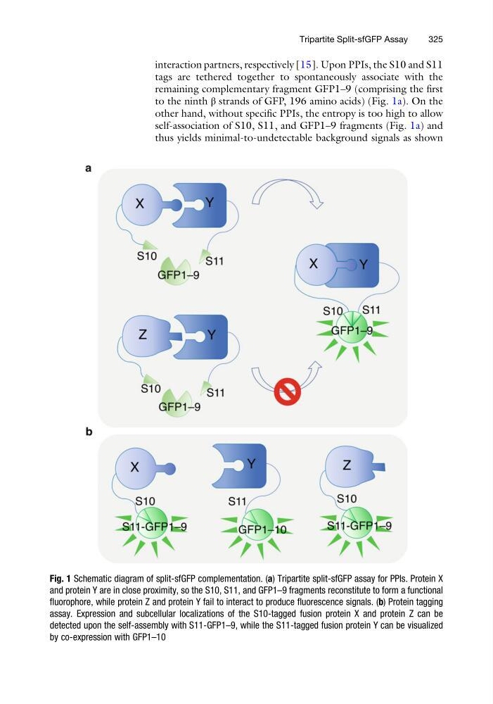
interaction partners, respectively [15]. Upon PPIs, the S10 and S11
tags are tethered together to spontaneously associate with the
remaining complementary fragment GFP1–9 (comprising the first
to the ninth β strands of GFP, 196 amino acids) (Fig. 1a). On the
other hand, without specific PPIs, the entropy is too high to allow
self-association of S10, S11, and GFP1–9 fragments (Fig. 1a) and
thus yields minimal-to-undetectable background signals as shown
Fig. 1 Schematic diagram of split-sfGFP complementation. (a) Tripartite split-sfGFP assay for PPIs. Protein X
and protein Y are in close proximity, so the S10, S11, and GFP1–9 fragments reconstitute to form a functional
fluorophore, while protein Z and protein Y fail to interact to produce fluorescence signals. (b) Protein tagging
assay. Expression and subcellular localizations of the S10-tagged fusion protein X and protein Z can be
detected upon the self-assembly with S11-GFP1–9, while the S11-tagged fusion protein Y can be visualized
by co-expression with GFP1–10
Tripartite Split-sfGFP Assay
325
tags are tethered together to spontaneously associate with the
remaining complementary fragment GFP1–9 (comprising the first
to the ninth β strands of GFP, 196 amino acids) (Fig. 1a). On the
other hand, without specific PPIs, the entropy is too high to allow
self-association of S10, S11, and GFP1–9 fragments (Fig. 1a) and
thus yields minimal-to-undetectable background signals as shown
Fig. 1 Schematic diagram of split-sfGFP complementation. (a) Tripartite split-sfGFP assay for PPIs. Protein X
and protein Y are in close proximity, so the S10, S11, and GFP1–9 fragments reconstitute to form a functional
fluorophore, while protein Z and protein Y fail to interact to produce fluorescence signals. (b) Protein tagging
assay. Expression and subcellular localizations of the S10-tagged fusion protein X and protein Z can be
detected upon the self-assembly with S11-GFP1–9, while the S11-tagged fusion protein Y can be visualized
by co-expression with GFP1–10
Tripartite Split-sfGFP Assay
325