

2.2 Microscope
Equipment
1. Inverted epifluorescence microscope ECLIPSE Ti2 (Nikon,
Tokyo, Japan) equipped with a microplate stage, 10x Plan
Apo (NA/0.45) lens (OFN25, Nikon), SPECTRA X Light
Engine
® (lumencor, OR, USA), Prime 95B sCMOS camera
(Photometrics, AZ, USA), filter sets, namely Dichroic mirror,
A
B
A-A ( 1 : 1 )
A
A
122.8
107
20
20
20
20
85.6
R 5
4 x
R 6
4 x
25
8
5
2
Rz
6.3
72
46
72
57
33
4xC0.5
C0.5
C0.5
C0.5
C0.5
C0.5
C0.5
C0.5
C0.5
C0.5
C0.5
7.9
6.8
25.4
32.9
26.3
19.8
2
C0.5
51.4
51.4
32.8
32.8
2
38
1
5
6
7
25
101
5
5
48
1.2
5
25
5
5
5
B
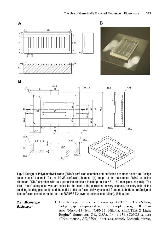
Fig. 3 Design of Polydimethylsiloxane (PDMS) perfusion chamber and perfusion chamber holder. (a) Design
schematic of the mold for the PDMS perfusion chamber. (b) Image of the assembled PDMS perfusion
chamber. PDMS chamber with four perfusion channels is sitting on the 48 Â 60 mm glass coverslip. The
three “dots” along each well are holes for the inlet of the perfusion delivery channel, an entry hole of the
seedling-holding pipette tip, and the outlet of the perfusion delivery channel from top to bottom. (c) Design of
the perfusion chamber holder for the ECRIPSE Ti2 inverted microscope (Nikon). Unit is mm
The Use of Genetically Encoded Fluorescent Biosensors
313
Equipment
1. Inverted epifluorescence microscope ECLIPSE Ti2 (Nikon,
Tokyo, Japan) equipped with a microplate stage, 10x Plan
Apo (NA/0.45) lens (OFN25, Nikon), SPECTRA X Light
Engine
® (lumencor, OR, USA), Prime 95B sCMOS camera
(Photometrics, AZ, USA), filter sets, namely Dichroic mirror,
A
B
A-A ( 1 : 1 )
A
A
122.8
107
20
20
20
20
85.6
R 5
4 x
R 6
4 x
25
8
5
2
Rz
6.3
72
46
72
57
33
4xC0.5
C0.5
C0.5
C0.5
C0.5
C0.5
C0.5
C0.5
C0.5
C0.5
C0.5
7.9
6.8
25.4
32.9
26.3
19.8
2
C0.5
51.4
51.4
32.8
32.8
2
38
1
5
6
7
25
101
5
5
48
1.2
5
25
5
5
5
B
Fig. 3 Design of Polydimethylsiloxane (PDMS) perfusion chamber and perfusion chamber holder. (a) Design
schematic of the mold for the PDMS perfusion chamber. (b) Image of the assembled PDMS perfusion
chamber. PDMS chamber with four perfusion channels is sitting on the 48 Â 60 mm glass coverslip. The
three “dots” along each well are holes for the inlet of the perfusion delivery channel, an entry hole of the
seedling-holding pipette tip, and the outlet of the perfusion delivery channel from top to bottom. (c) Design of
the perfusion chamber holder for the ECRIPSE Ti2 inverted microscope (Nikon). Unit is mm
The Use of Genetically Encoded Fluorescent Biosensors
313