

3.3.2 Analysis and
Sorting of Nuclei using the
S3
1. For analysis and sorting of PI-stained nuclei using the threelaser system, create panel templates as follows: a bivariate histogram of SSC vs. log FL3(A) [586/25], a bivariate plot of FSC
(A) vs. FSC(H) to eliminate nuclear doublets (a consequence
of inherent nuclear stickiness), a bivariate template of log FL3
(A) vs. time, to verify staining stability, and a univariate plot of
FL3(A) log. If the nuclei are from transgenic plants and are
accumulating GFP, add to the above a bivariate plot of
SSC vs. log FL2(A) [525/30 nm], a bivariate plot of log FL2
(A) vs. log FL3(A), and a univariate plot of log FL2(A).
2. Validate the cytometer using calibration beads or a nuclear
suspension obtained from recommended controls (a leaf of
pea, maize, tomato, or Arabidopsis thaliana stained with PI
in the presence of RNAase). For cell cycle analysis of
non-endoreduplicative species, you can set the FL3 channel
to linear amplification. For endoreduplication measurements
(Arabidopsis, etc.), logarithmic amplification is required, using
FL3 [586/25] for detection of the fluorescence signals from
PI. Analysis of the homogenate, based on light scatter and
fluorescence signals produced by excitation by all of the lasers
should be triggered by the PI channel fluorescence (FL3
[586/25]), in contrast to the usual practice of triggering
using FALS).
3. Perform analysis eliminating, via gating, any nonspecific signals
coming from debris, aggregated nuclei, etc. For this purpose,
gating on single nuclei, as defined along the diagonal of the
FSC (A) vs. FSC (H) plot, can be useful since only the gated
signals representing single nuclei are then included in the
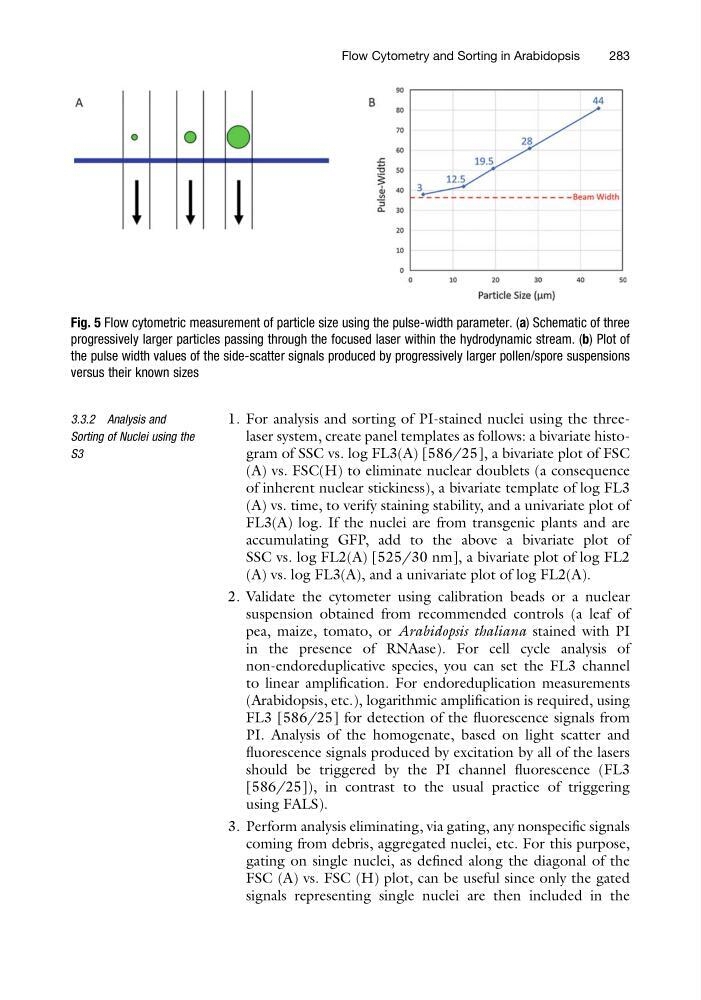
Fig. 5 Flow cytometric measurement of particle size using the pulse-width parameter. (a) Schematic of three
progressively larger particles passing through the focused laser within the hydrodynamic stream. (b) Plot of
the pulse width values of the side-scatter signals produced by progressively larger pollen/spore suspensions
versus their known sizes
Flow Cytometry and Sorting in Arabidopsis
283
Sorting of Nuclei using the
S3
1. For analysis and sorting of PI-stained nuclei using the threelaser system, create panel templates as follows: a bivariate histogram of SSC vs. log FL3(A) [586/25], a bivariate plot of FSC
(A) vs. FSC(H) to eliminate nuclear doublets (a consequence
of inherent nuclear stickiness), a bivariate template of log FL3
(A) vs. time, to verify staining stability, and a univariate plot of
FL3(A) log. If the nuclei are from transgenic plants and are
accumulating GFP, add to the above a bivariate plot of
SSC vs. log FL2(A) [525/30 nm], a bivariate plot of log FL2
(A) vs. log FL3(A), and a univariate plot of log FL2(A).
2. Validate the cytometer using calibration beads or a nuclear
suspension obtained from recommended controls (a leaf of
pea, maize, tomato, or Arabidopsis thaliana stained with PI
in the presence of RNAase). For cell cycle analysis of
non-endoreduplicative species, you can set the FL3 channel
to linear amplification. For endoreduplication measurements
(Arabidopsis, etc.), logarithmic amplification is required, using
FL3 [586/25] for detection of the fluorescence signals from
PI. Analysis of the homogenate, based on light scatter and
fluorescence signals produced by excitation by all of the lasers
should be triggered by the PI channel fluorescence (FL3
[586/25]), in contrast to the usual practice of triggering
using FALS).
3. Perform analysis eliminating, via gating, any nonspecific signals
coming from debris, aggregated nuclei, etc. For this purpose,
gating on single nuclei, as defined along the diagonal of the
FSC (A) vs. FSC (H) plot, can be useful since only the gated
signals representing single nuclei are then included in the
Fig. 5 Flow cytometric measurement of particle size using the pulse-width parameter. (a) Schematic of three
progressively larger particles passing through the focused laser within the hydrodynamic stream. (b) Plot of
the pulse width values of the side-scatter signals produced by progressively larger pollen/spore suspensions
versus their known sizes
Flow Cytometry and Sorting in Arabidopsis
283