

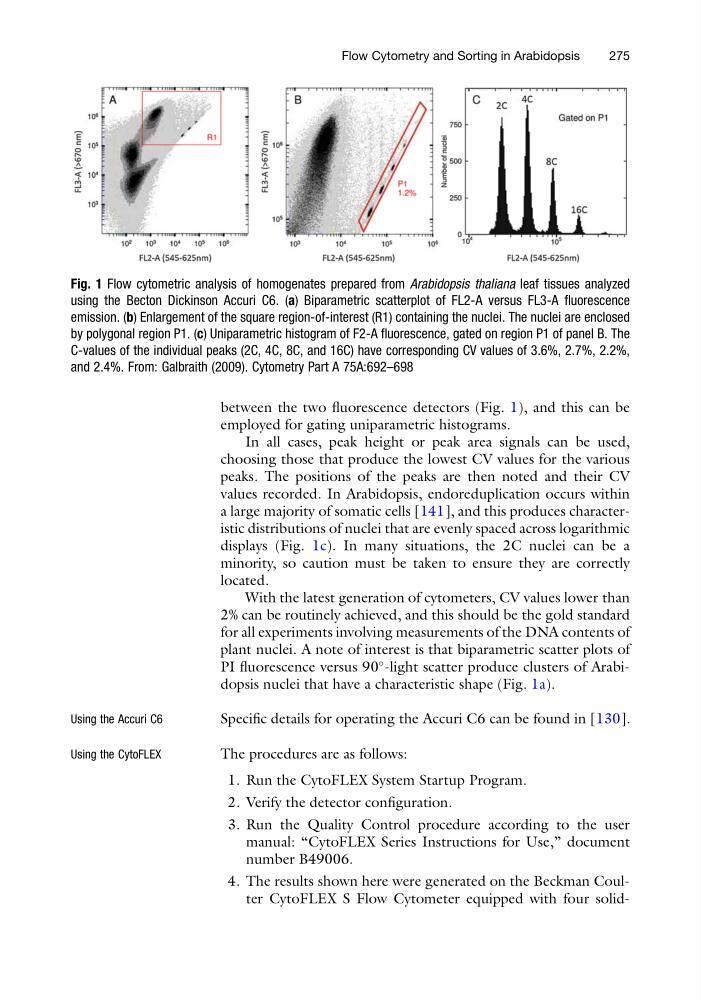
between the two fluorescence detectors (Fig. 1), and this can be
employed for gating uniparametric histograms.
In all cases, peak height or peak area signals can be used,
choosing those that produce the lowest CV values for the various
peaks. The positions of the peaks are then noted and their CV
values recorded. In Arabidopsis, endoreduplication occurs within
a large majority of somatic cells [141], and this produces characteristic distributions of nuclei that are evenly spaced across logarithmic
displays (Fig. 1c). In many situations, the 2C nuclei can be a
minority, so caution must be taken to ensure they are correctly
located.
With the latest generation of cytometers, CV values lower than
2% can be routinely achieved, and this should be the gold standard
for all experiments involving measurements of the DNA contents of
plant nuclei. A note of interest is that biparametric scatter plots of
PI fluorescence versus 90
-light scatter produce clusters of Arabidopsis nuclei that have a characteristic shape (Fig. 1a).
Using the Accuri C6
Specific details for operating the Accuri C6 can be found in [130].
Using the CytoFLEX
The procedures are as follows:
1. Run the CytoFLEX System Startup Program.
2. Verify the detector configuration.
3. Run the Quality Control procedure according to the user
manual: “CytoFLEX Series Instructions for Use,” document
number B49006.
4. The results shown here were generated on the Beckman Coulter CytoFLEX S Flow Cytometer equipped with four solidFig. 1 Flow cytometric analysis of homogenates prepared from Arabidopsis thaliana leaf tissues analyzed
using the Becton Dickinson Accuri C6. (a) Biparametric scatterplot of FL2-A versus FL3-A fluorescence
emission. (b) Enlargement of the square region-of-interest (R1) containing the nuclei. The nuclei are enclosed
by polygonal region P1. (c) Uniparametric histogram of F2-A fluorescence, gated on region P1 of panel B. The
C-values of the individual peaks (2C, 4C, 8C, and 16C) have corresponding CV values of 3.6%, 2.7%, 2.2%,
and 2.4%. From: Galbraith (2009). Cytometry Part A 75A:692–698
Flow Cytometry and Sorting in Arabidopsis
275
employed for gating uniparametric histograms.
In all cases, peak height or peak area signals can be used,
choosing those that produce the lowest CV values for the various
peaks. The positions of the peaks are then noted and their CV
values recorded. In Arabidopsis, endoreduplication occurs within
a large majority of somatic cells [141], and this produces characteristic distributions of nuclei that are evenly spaced across logarithmic
displays (Fig. 1c). In many situations, the 2C nuclei can be a
minority, so caution must be taken to ensure they are correctly
located.
With the latest generation of cytometers, CV values lower than
2% can be routinely achieved, and this should be the gold standard
for all experiments involving measurements of the DNA contents of
plant nuclei. A note of interest is that biparametric scatter plots of
PI fluorescence versus 90
-light scatter produce clusters of Arabidopsis nuclei that have a characteristic shape (Fig. 1a).
Using the Accuri C6
Specific details for operating the Accuri C6 can be found in [130].
Using the CytoFLEX
The procedures are as follows:
1. Run the CytoFLEX System Startup Program.
2. Verify the detector configuration.
3. Run the Quality Control procedure according to the user
manual: “CytoFLEX Series Instructions for Use,” document
number B49006.
4. The results shown here were generated on the Beckman Coulter CytoFLEX S Flow Cytometer equipped with four solidFig. 1 Flow cytometric analysis of homogenates prepared from Arabidopsis thaliana leaf tissues analyzed
using the Becton Dickinson Accuri C6. (a) Biparametric scatterplot of FL2-A versus FL3-A fluorescence
emission. (b) Enlargement of the square region-of-interest (R1) containing the nuclei. The nuclei are enclosed
by polygonal region P1. (c) Uniparametric histogram of F2-A fluorescence, gated on region P1 of panel B. The
C-values of the individual peaks (2C, 4C, 8C, and 16C) have corresponding CV values of 3.6%, 2.7%, 2.2%,
and 2.4%. From: Galbraith (2009). Cytometry Part A 75A:692–698
Flow Cytometry and Sorting in Arabidopsis
275