

that was used to purify the size markers after
32 P-labeling. Wrap
the gel in plastic film and cover it with a phosphor screen and
keep them in dark for 30 min.
9. Expose the phosphor screen and print out the image as the
original size (Fig. 3a). Align the gel on glass to the paper
printout and then use a clean blade to cut the gel framed by
32 P-labeled 19 to 24-nt markers. Chop that gel slices into small
pieces and carefully transfer them to a 1.5 mL tube with 350 μL
0.4 M NaCl (DEPC treated). Use clean water to rinse and kim
wipes to clean the blades between samples.
10. Tumble in the Eppendorf thermomixer at a speed of
1200 rpm/min at 4
C overnight.
11. Centrifuge at 12,000 g for 15 min at 4
C. Take supernatant
out to a new tube. Add 100 μL 0.4 M NaCl (DEPC treated) to
each old tube and tumble 30 min at 4
C.
12. Centrifuge at 12,000 g for 5 min at 4
C. Take the supernatants
out to the new tubes and add 1 mL ethanol and 1 μL glycogen,
mix well and incubate at À80
C for at least 1 h or À20
C
overnight.
13. Centrifuge at maximum speed for 15 min at 4
C. Discard
supernatant.
14. Wash the pellet with 500 μL 75% ethanol. Then centrifuge at
13800 g for 10 min. Discard ethanol. Spin down quickly and
carefully remove the residual ethanol.
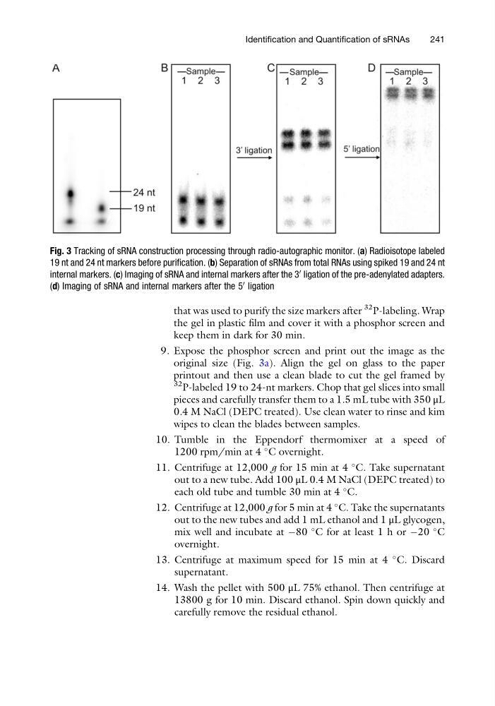
Fig. 3 Tracking of sRNA construction processing through radio-autographic monitor. (a) Radioisotope labeled
19 nt and 24 nt markers before purification. (b) Separation of sRNAs from total RNAs using spiked 19 and 24 nt
internal markers. (c) Imaging of sRNA and internal markers after the 3
0
ligation of the pre-adenylated adapters.
(d) Imaging of sRNA and internal markers after the 5
0
ligation
Identification and Quantification of sRNAs
241
32 P-labeling. Wrap
the gel in plastic film and cover it with a phosphor screen and
keep them in dark for 30 min.
9. Expose the phosphor screen and print out the image as the
original size (Fig. 3a). Align the gel on glass to the paper
printout and then use a clean blade to cut the gel framed by
32 P-labeled 19 to 24-nt markers. Chop that gel slices into small
pieces and carefully transfer them to a 1.5 mL tube with 350 μL
0.4 M NaCl (DEPC treated). Use clean water to rinse and kim
wipes to clean the blades between samples.
10. Tumble in the Eppendorf thermomixer at a speed of
1200 rpm/min at 4
C overnight.
11. Centrifuge at 12,000 g for 15 min at 4
C. Take supernatant
out to a new tube. Add 100 μL 0.4 M NaCl (DEPC treated) to
each old tube and tumble 30 min at 4
C.
12. Centrifuge at 12,000 g for 5 min at 4
C. Take the supernatants
out to the new tubes and add 1 mL ethanol and 1 μL glycogen,
mix well and incubate at À80
C for at least 1 h or À20
C
overnight.
13. Centrifuge at maximum speed for 15 min at 4
C. Discard
supernatant.
14. Wash the pellet with 500 μL 75% ethanol. Then centrifuge at
13800 g for 10 min. Discard ethanol. Spin down quickly and
carefully remove the residual ethanol.
Fig. 3 Tracking of sRNA construction processing through radio-autographic monitor. (a) Radioisotope labeled
19 nt and 24 nt markers before purification. (b) Separation of sRNAs from total RNAs using spiked 19 and 24 nt
internal markers. (c) Imaging of sRNA and internal markers after the 3
0
ligation of the pre-adenylated adapters.
(d) Imaging of sRNA and internal markers after the 5
0
ligation
Identification and Quantification of sRNAs
241