

3. Excise the cotyledons and the hypocotyl, then transfer them
separately in two different Petri dishes containing CIM for
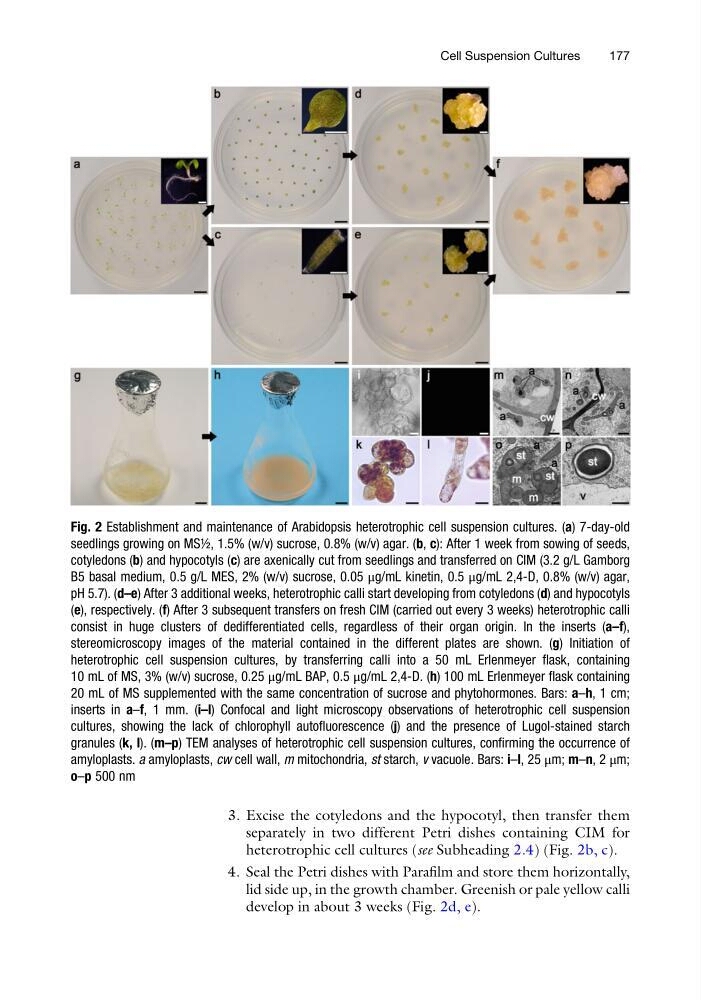
heterotrophic cell cultures (see Subheading 2.4) (Fig. 2b, c).
4. Seal the Petri dishes with Parafilm and store them horizontally,
lid side up, in the growth chamber. Greenish or pale yellow calli
develop in about 3 weeks (Fig. 2d, e).
Fig. 2 Establishment and maintenance of Arabidopsis heterotrophic cell suspension cultures. (a) 7-day-old
seedlings growing on MS½, 1.5% (w/v) sucrose, 0.8% (w/v) agar. (b, c): After 1 week from sowing of seeds,
cotyledons (b) and hypocotyls (c) are axenically cut from seedlings and transferred on CIM (3.2 g/L Gamborg
B5 basal medium, 0.5 g/L MES, 2% (w/v) sucrose, 0.05 μg/mL kinetin, 0.5 μg/mL 2,4-D, 0.8% (w/v) agar,
pH 5.7). (d–e) After 3 additional weeks, heterotrophic calli start developing from cotyledons (d) and hypocotyls
(e), respectively. (f) After 3 subsequent transfers on fresh CIM (carried out every 3 weeks) heterotrophic calli
consist in huge clusters of dedifferentiated cells, regardless of their organ origin. In the inserts (a–f),
stereomicroscopy images of the material contained in the different plates are shown. (g) Initiation of
heterotrophic cell suspension cultures, by transferring calli into a 50 mL Erlenmeyer flask, containing
10 mL of MS, 3% (w/v) sucrose, 0.25 μg/mL BAP, 0.5 μg/mL 2,4-D. (h) 100 mL Erlenmeyer flask containing
20 mL of MS supplemented with the same concentration of sucrose and phytohormones. Bars: a–h, 1 cm;
inserts in a–f, 1 mm. (i–l) Confocal and light microscopy observations of heterotrophic cell suspension
cultures, showing the lack of chlorophyll autofluorescence (j) and the presence of Lugol-stained starch
granules (k, l). (m–p) TEM analyses of heterotrophic cell suspension cultures, confirming the occurrence of
amyloplasts. a amyloplasts, cw cell wall, m mitochondria, st starch, v vacuole. Bars: i–l, 25 μm; m–n, 2 μm;
o–p 500 nm
Cell Suspension Cultures
177
separately in two different Petri dishes containing CIM for
heterotrophic cell cultures (see Subheading 2.4) (Fig. 2b, c).
4. Seal the Petri dishes with Parafilm and store them horizontally,
lid side up, in the growth chamber. Greenish or pale yellow calli
develop in about 3 weeks (Fig. 2d, e).
Fig. 2 Establishment and maintenance of Arabidopsis heterotrophic cell suspension cultures. (a) 7-day-old
seedlings growing on MS½, 1.5% (w/v) sucrose, 0.8% (w/v) agar. (b, c): After 1 week from sowing of seeds,
cotyledons (b) and hypocotyls (c) are axenically cut from seedlings and transferred on CIM (3.2 g/L Gamborg
B5 basal medium, 0.5 g/L MES, 2% (w/v) sucrose, 0.05 μg/mL kinetin, 0.5 μg/mL 2,4-D, 0.8% (w/v) agar,
pH 5.7). (d–e) After 3 additional weeks, heterotrophic calli start developing from cotyledons (d) and hypocotyls
(e), respectively. (f) After 3 subsequent transfers on fresh CIM (carried out every 3 weeks) heterotrophic calli
consist in huge clusters of dedifferentiated cells, regardless of their organ origin. In the inserts (a–f),
stereomicroscopy images of the material contained in the different plates are shown. (g) Initiation of
heterotrophic cell suspension cultures, by transferring calli into a 50 mL Erlenmeyer flask, containing
10 mL of MS, 3% (w/v) sucrose, 0.25 μg/mL BAP, 0.5 μg/mL 2,4-D. (h) 100 mL Erlenmeyer flask containing
20 mL of MS supplemented with the same concentration of sucrose and phytohormones. Bars: a–h, 1 cm;
inserts in a–f, 1 mm. (i–l) Confocal and light microscopy observations of heterotrophic cell suspension
cultures, showing the lack of chlorophyll autofluorescence (j) and the presence of Lugol-stained starch
granules (k, l). (m–p) TEM analyses of heterotrophic cell suspension cultures, confirming the occurrence of
amyloplasts. a amyloplasts, cw cell wall, m mitochondria, st starch, v vacuole. Bars: i–l, 25 μm; m–n, 2 μm;
o–p 500 nm
Cell Suspension Cultures
177