

4. With a sterile scalpel remove the residues of
non-dedifferentiated cotyledons and cut the green calli in two
or four pieces (see Note 14).
5. With sterile forceps carefully transfer the material deriving from
about ten calli into the 50 mL Erlenmeyer flask containing the
liquid culture medium (see Note 15).
6. Reduce the dimension of the aluminum foil lid by using scissors, taking care to preserve sterility during this operation (see
Note 16).
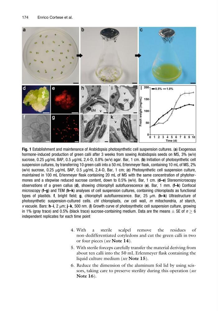
Fig. 1 Establishment and maintenance of Arabidopsis photosynthetic cell suspension cultures. (a) Exogenous
hormone-induced production of green calli after 3 weeks from sowing Arabidopsis seeds on MS, 3% (w/v)
sucrose, 0.25 μg/mL BAP, 0.5 μg/mL 2,4-D, 0.8% (w/v) agar. Bar, 1 cm. (b) Initiation of photosynthetic cell
suspension cultures, by transferring 10 green calli into a 50 mL Erlenmeyer flask, containing 10 mL of MS, 2%
(w/v) sucrose, 0.25 μg/mL BAP, 0.5 μg/mL 2,4-D, Bar, 1 cm; (c) Photosynthetic cell suspension culture,
maintained in 100 mL Erlenmeyer flask containing 20 mL of MS with the same concentration of phytohormones and a stepwise reduced sucrose content, down to 0.5% (w/v). Bar, 1 cm. (d–e) Stereomicroscopy
observations of a green callus (d), showing chlorophyll autofluorescence (e). Bar, 1 mm. (f–k) Confocal
microscopy (f–g) and TEM (h–k) analyses of cell suspension cultures, containing chloroplasts as functional
types of plastids. f, bright field; g, chlorophyll autofluorescence. Bar, 25 μm. (h–k) Ultrastructure of
photosynthetic suspension-cultured cells. chl chloroplasts, cw cell wall, m mitochondria, st starch,
v vacuole. Bars: h–i, 2 μm; j–k, 500 nm. (l) Growth curve of photosynthetic cell suspension culture, growing
in 1% (gray trace) and 0.5% (black trace) sucrose-containing medium. Data are the means Æ SE of n ! 6
independent replicates for each time point
174
Enrico Cortese et al.
non-dedifferentiated cotyledons and cut the green calli in two
or four pieces (see Note 14).
5. With sterile forceps carefully transfer the material deriving from
about ten calli into the 50 mL Erlenmeyer flask containing the
liquid culture medium (see Note 15).
6. Reduce the dimension of the aluminum foil lid by using scissors, taking care to preserve sterility during this operation (see
Note 16).
Fig. 1 Establishment and maintenance of Arabidopsis photosynthetic cell suspension cultures. (a) Exogenous
hormone-induced production of green calli after 3 weeks from sowing Arabidopsis seeds on MS, 3% (w/v)
sucrose, 0.25 μg/mL BAP, 0.5 μg/mL 2,4-D, 0.8% (w/v) agar. Bar, 1 cm. (b) Initiation of photosynthetic cell
suspension cultures, by transferring 10 green calli into a 50 mL Erlenmeyer flask, containing 10 mL of MS, 2%
(w/v) sucrose, 0.25 μg/mL BAP, 0.5 μg/mL 2,4-D, Bar, 1 cm; (c) Photosynthetic cell suspension culture,
maintained in 100 mL Erlenmeyer flask containing 20 mL of MS with the same concentration of phytohormones and a stepwise reduced sucrose content, down to 0.5% (w/v). Bar, 1 cm. (d–e) Stereomicroscopy
observations of a green callus (d), showing chlorophyll autofluorescence (e). Bar, 1 mm. (f–k) Confocal
microscopy (f–g) and TEM (h–k) analyses of cell suspension cultures, containing chloroplasts as functional
types of plastids. f, bright field; g, chlorophyll autofluorescence. Bar, 25 μm. (h–k) Ultrastructure of
photosynthetic suspension-cultured cells. chl chloroplasts, cw cell wall, m mitochondria, st starch,
v vacuole. Bars: h–i, 2 μm; j–k, 500 nm. (l) Growth curve of photosynthetic cell suspension culture, growing
in 1% (gray trace) and 0.5% (black trace) sucrose-containing medium. Data are the means Æ SE of n ! 6
independent replicates for each time point
174
Enrico Cortese et al.