

3 Methods
3.1 Protoplast
Isolation
1. Transfer 50 mL 5-day-old Arabidopsis PSBD cell suspension
culture into 50 mL conical tubes in a laminar flow hood.
2. Pellet down cells by centrifugation at 100 Â g for 5 min at
room temperature, then remove the supernatant.
3. Sterilize 50 mL freshly prepared enzyme solution using
0.22 μm filter and transfer the solution into the tubes with
the pelleted down cells to a final 50 mL volume. Slowly invert
several times for mixing.
4. Transfer 50 mL well-mixed culture cells into sterilized Erlenmeyer flask and digest Arabidopsis cultured cells at 27
C,
65 rpm shaking for 1.5–2 h. Check the protoplasts under
light microscope. For the good digestion, more than 95%
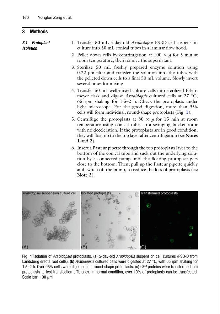
cells will form individual, round-shape protoplasts (Fig. 1).
5. Centrifuge the protoplasts at 80 Â g for 15 min at room
temperature using conical tubes in a swinging bucket rotor
with no deceleration. If the protoplasts are in good condition,
they will float up to the top layer after centrifugation (see Notes
1 and 2).
6. Insert a Pasteur pipette through the top protoplasts layer to the
bottom of the conical tube and suck out the underlying solution by a connected pump until the floating protoplast gets
close to the bottom. Then, pull up the Pasteur pipette quickly
and switch off the pump, to reduce the loss of protoplasts (see
Note 3).
Fig. 1 Isolation of Arabidopsis protoplasts. (a) 5-day-old Arabidopsis suspension cell cultures (PSB-D from
Landsberg erecta root cells). (b) Arabidopsis cultured cells were digested at 27
C, with 65 rpm shaking for
1.5–2 h. Over 95% cells were digested into round-shape protoplasts. (c) GFP proteins were transformed into
protoplasts to test transfection efficiency. In normal condition, over 10% of protoplasts can be transfected.
Scale bar, 100 μm
160
Yonglun Zeng et al.
3.1 Protoplast
Isolation
1. Transfer 50 mL 5-day-old Arabidopsis PSBD cell suspension
culture into 50 mL conical tubes in a laminar flow hood.
2. Pellet down cells by centrifugation at 100 Â g for 5 min at
room temperature, then remove the supernatant.
3. Sterilize 50 mL freshly prepared enzyme solution using
0.22 μm filter and transfer the solution into the tubes with
the pelleted down cells to a final 50 mL volume. Slowly invert
several times for mixing.
4. Transfer 50 mL well-mixed culture cells into sterilized Erlenmeyer flask and digest Arabidopsis cultured cells at 27
C,
65 rpm shaking for 1.5–2 h. Check the protoplasts under
light microscope. For the good digestion, more than 95%
cells will form individual, round-shape protoplasts (Fig. 1).
5. Centrifuge the protoplasts at 80 Â g for 15 min at room
temperature using conical tubes in a swinging bucket rotor
with no deceleration. If the protoplasts are in good condition,
they will float up to the top layer after centrifugation (see Notes
1 and 2).
6. Insert a Pasteur pipette through the top protoplasts layer to the
bottom of the conical tube and suck out the underlying solution by a connected pump until the floating protoplast gets
close to the bottom. Then, pull up the Pasteur pipette quickly
and switch off the pump, to reduce the loss of protoplasts (see
Note 3).
Fig. 1 Isolation of Arabidopsis protoplasts. (a) 5-day-old Arabidopsis suspension cell cultures (PSB-D from
Landsberg erecta root cells). (b) Arabidopsis cultured cells were digested at 27
C, with 65 rpm shaking for
1.5–2 h. Over 95% cells were digested into round-shape protoplasts. (c) GFP proteins were transformed into
protoplasts to test transfection efficiency. In normal condition, over 10% of protoplasts can be transfected.
Scale bar, 100 μm
160
Yonglun Zeng et al.