

6. Take the coverslip with the forceps and rinse it in the beaker.
Place the coverslip back in the IF chamber and incubate with
the coverslip with 190 μL of primary antibody (mouse antialpha-tubulin, 1:1000) in IF solution for 45 min at RT (see
Note 13).
7. Wash the coverslips three times with 190 μL PBS for 5 min
at RT.
8. Incubate with 190 μL of secondary antibody (goat anti-mouse
Alexa 647, 1:1500) in PBS 1Â and BSA 0.1% for 45 min at RT
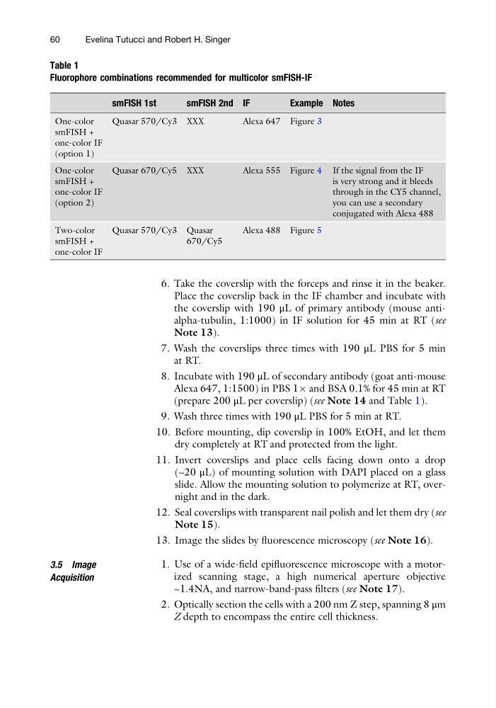
(prepare 200 μL per coverslip) (see Note 14 and Table 1).
9. Wash three times with 190 μL PBS for 5 min at RT.
10. Before mounting, dip coverslip in 100% EtOH, and let them
dry completely at RT and protected from the light.
11. Invert coverslips and place cells facing down onto a drop
(~20 μL) of mounting solution with DAPI placed on a glass
slide. Allow the mounting solution to polymerize at RT, overnight and in the dark.
12. Seal coverslips with transparent nail polish and let them dry (see
Note 15).
13. Image the slides by fluorescence microscopy (see Note 16).
3.5 Image
Acquisition
1. Use of a wide-field epifluorescence microscope with a motorized scanning stage, a high numerical aperture objective
~1.4NA, and narrow-band-pass filters (see Note 17).
2. Optically section the cells with a 200 nm Z step, spanning 8 μm
Z depth to encompass the entire cell thickness.
Table 1
Fluorophore combinations recommended for multicolor smFISH-IF
smFISH 1st
smFISH 2nd IF
Example Notes
One-color
smFISH +
one-color IF
(option 1)
Quasar 570/Cy3 XXX
Alexa 647 Figure 3
One-color
smFISH +
one-color IF
(option 2)
Quasar 670/Cy5 XXX
Alexa 555 Figure 4 If the signal from the IF
is very strong and it bleeds
through in the CY5 channel,
you can use a secondary
conjugated with Alexa 488
Two-color
smFISH +
one-color IF
Quasar 570/Cy3 Quasar
670/Cy5
Alexa 488 Figure 5
60
Evelina Tutucci and Robert H. Singer
Place the coverslip back in the IF chamber and incubate with
the coverslip with 190 μL of primary antibody (mouse antialpha-tubulin, 1:1000) in IF solution for 45 min at RT (see
Note 13).
7. Wash the coverslips three times with 190 μL PBS for 5 min
at RT.
8. Incubate with 190 μL of secondary antibody (goat anti-mouse
Alexa 647, 1:1500) in PBS 1Â and BSA 0.1% for 45 min at RT
(prepare 200 μL per coverslip) (see Note 14 and Table 1).
9. Wash three times with 190 μL PBS for 5 min at RT.
10. Before mounting, dip coverslip in 100% EtOH, and let them
dry completely at RT and protected from the light.
11. Invert coverslips and place cells facing down onto a drop
(~20 μL) of mounting solution with DAPI placed on a glass
slide. Allow the mounting solution to polymerize at RT, overnight and in the dark.
12. Seal coverslips with transparent nail polish and let them dry (see
Note 15).
13. Image the slides by fluorescence microscopy (see Note 16).
3.5 Image
Acquisition
1. Use of a wide-field epifluorescence microscope with a motorized scanning stage, a high numerical aperture objective
~1.4NA, and narrow-band-pass filters (see Note 17).
2. Optically section the cells with a 200 nm Z step, spanning 8 μm
Z depth to encompass the entire cell thickness.
Table 1
Fluorophore combinations recommended for multicolor smFISH-IF
smFISH 1st
smFISH 2nd IF
Example Notes
One-color
smFISH +
one-color IF
(option 1)
Quasar 570/Cy3 XXX
Alexa 647 Figure 3
One-color
smFISH +
one-color IF
(option 2)
Quasar 670/Cy5 XXX
Alexa 555 Figure 4 If the signal from the IF
is very strong and it bleeds
through in the CY5 channel,
you can use a secondary
conjugated with Alexa 488
Two-color
smFISH +
one-color IF
Quasar 570/Cy3 Quasar
670/Cy5
Alexa 488 Figure 5
60
Evelina Tutucci and Robert H. Singer