

expression vector (or the pK7WG2D empty vector)-encoded
GFP runs at ~33 kDa (see Fig. 1b). Take multiple pictures of
the membrane with various exposure times. Be sure to have
images where signal intensities are not at saturating levels. Use
these images for accurate quantitation of the protein bands.
8. Use your imaging software (e.g., Image Lab) for identification
and accurate quantitation of protein bands. Identification of
bands can be done automatically or manually. Manual labeling
of the bands is necessary if they are too faint for automatic
detection. Image Lab generates an analysis table, which contains absolute values of band intensities (with background
subtracted). The analysis table can be exported to Excel for
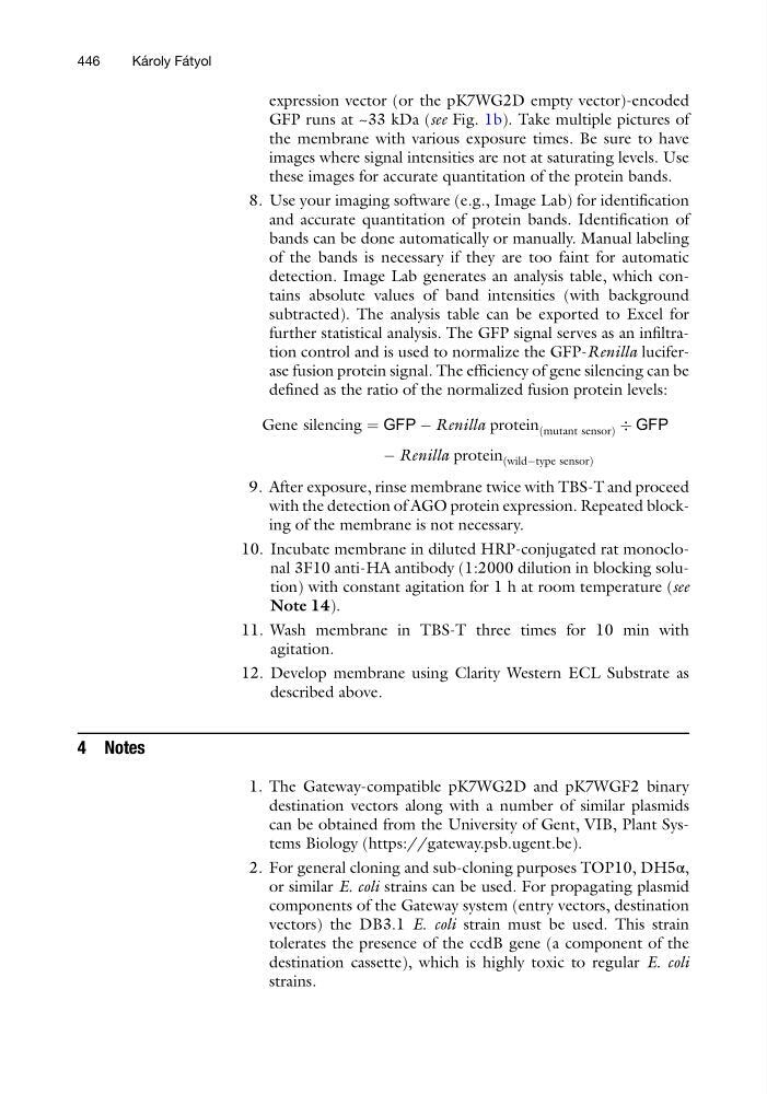
further statistical analysis. The GFP signal serves as an infiltration control and is used to normalize the GFP-Renilla luciferase fusion protein signal. The efficiency of gene silencing can be
defined as the ratio of the normalized fusion protein levels:
Gene silencing ¼ GFP À Renilla protein mutant sensor
ð
Þ Ä GFP
À Renilla protein wildÀtype sensor
ð
Þ
9. After exposure, rinse membrane twice with TBS-T and proceed
with the detection of AGO protein expression. Repeated blocking of the membrane is not necessary.
10. Incubate membrane in diluted HRP-conjugated rat monoclonal 3F10 anti-HA antibody (1:2000 dilution in blocking solution) with constant agitation for 1 h at room temperature (see
Note 14).
11. Wash membrane in TBS-T three times for 10 min with
agitation.
12. Develop membrane using Clarity Western ECL Substrate as
described above.
4 Notes
1. The Gateway-compatible pK7WG2D and pK7WGF2 binary
destination vectors along with a number of similar plasmids
can be obtained from the University of Gent, VIB, Plant Systems Biology (https://gateway.psb.ugent.be).
2. For general cloning and sub-cloning purposes TOP10, DH5α,
or similar E. coli strains can be used. For propagating plasmid
components of the Gateway system (entry vectors, destination
vectors) the DB3.1 E. coli strain must be used. This strain
tolerates the presence of the ccdB gene (a component of the
destination cassette), which is highly toxic to regular E. coli
strains.
446
Ka ´ roly Fa ´ tyol
GFP runs at ~33 kDa (see Fig. 1b). Take multiple pictures of
the membrane with various exposure times. Be sure to have
images where signal intensities are not at saturating levels. Use
these images for accurate quantitation of the protein bands.
8. Use your imaging software (e.g., Image Lab) for identification
and accurate quantitation of protein bands. Identification of
bands can be done automatically or manually. Manual labeling
of the bands is necessary if they are too faint for automatic
detection. Image Lab generates an analysis table, which contains absolute values of band intensities (with background
subtracted). The analysis table can be exported to Excel for
further statistical analysis. The GFP signal serves as an infiltration control and is used to normalize the GFP-Renilla luciferase fusion protein signal. The efficiency of gene silencing can be
defined as the ratio of the normalized fusion protein levels:
Gene silencing ¼ GFP À Renilla protein mutant sensor
ð
Þ Ä GFP
À Renilla protein wildÀtype sensor
ð
Þ
9. After exposure, rinse membrane twice with TBS-T and proceed
with the detection of AGO protein expression. Repeated blocking of the membrane is not necessary.
10. Incubate membrane in diluted HRP-conjugated rat monoclonal 3F10 anti-HA antibody (1:2000 dilution in blocking solution) with constant agitation for 1 h at room temperature (see
Note 14).
11. Wash membrane in TBS-T three times for 10 min with
agitation.
12. Develop membrane using Clarity Western ECL Substrate as
described above.
4 Notes
1. The Gateway-compatible pK7WG2D and pK7WGF2 binary
destination vectors along with a number of similar plasmids
can be obtained from the University of Gent, VIB, Plant Systems Biology (https://gateway.psb.ugent.be).
2. For general cloning and sub-cloning purposes TOP10, DH5α,
or similar E. coli strains can be used. For propagating plasmid
components of the Gateway system (entry vectors, destination
vectors) the DB3.1 E. coli strain must be used. This strain
tolerates the presence of the ccdB gene (a component of the
destination cassette), which is highly toxic to regular E. coli
strains.
446
Ka ´ roly Fa ´ tyol