

2. Pipette out the PBS and add 250 μL 4% PFA pre-warmed to
37
C per well. Incubate at RT for 30 min to fix the cells.
3. Wash the cells with PBS briefly, and then permeabilize cells
with 0.5% TritonX-100 for 30 min.
4. Wash the cells with 0.05% TritonX-100 for 5 min, followed by
incubation for 60 min in blocking buffer.
5. Stain the cells with human anti-centromere antibodies (final
concentration ¼ 4.2 μg/mL) in blocking buffer for 60 min.
6. Wash cells thrice with 0.05% TritonX-100 for 5 min, and then
stain cells with Alexa647-labeled anti-human secondary antibodies (final concentration ¼ 1 μg/mL) in blocking buffer for
60 min.
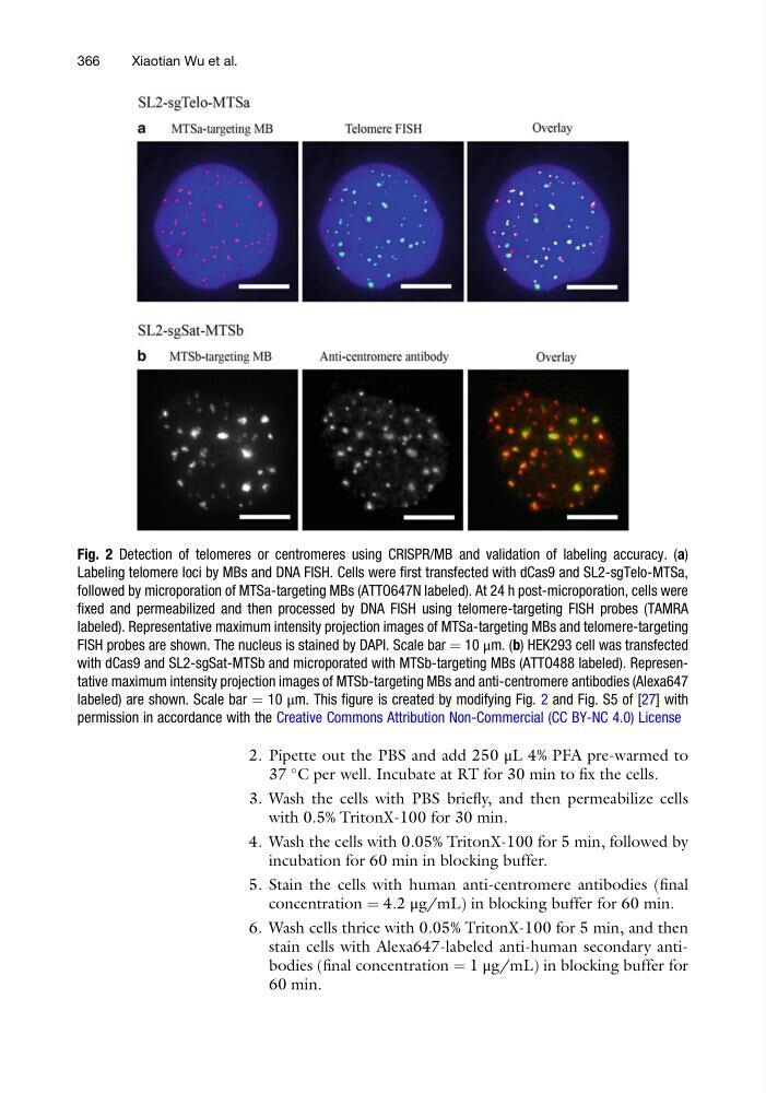
Fig. 2 Detection of telomeres or centromeres using CRISPR/MB and validation of labeling accuracy. (a)
Labeling telomere loci by MBs and DNA FISH. Cells were first transfected with dCas9 and SL2-sgTelo-MTSa,
followed by microporation of MTSa-targeting MBs (ATTO647N labeled). At 24 h post-microporation, cells were
fixed and permeabilized and then processed by DNA FISH using telomere-targeting FISH probes (TAMRA
labeled). Representative maximum intensity projection images of MTSa-targeting MBs and telomere-targeting
FISH probes are shown. The nucleus is stained by DAPI. Scale bar ¼ 10 μm. (b) HEK293 cell was transfected
with dCas9 and SL2-sgSat-MTSb and microporated with MTSb-targeting MBs (ATTO488 labeled). Representative maximum intensity projection images of MTSb-targeting MBs and anti-centromere antibodies (Alexa647
labeled) are shown. Scale bar ¼ 10 μm. This figure is created by modifying Fig. 2 and Fig. S5 of [27] with
permission in accordance with the Creative Commons Attribution Non-Commercial (CC BY-NC 4.0) License
366
Xiaotian Wu et al.
37
C per well. Incubate at RT for 30 min to fix the cells.
3. Wash the cells with PBS briefly, and then permeabilize cells
with 0.5% TritonX-100 for 30 min.
4. Wash the cells with 0.05% TritonX-100 for 5 min, followed by
incubation for 60 min in blocking buffer.
5. Stain the cells with human anti-centromere antibodies (final
concentration ¼ 4.2 μg/mL) in blocking buffer for 60 min.
6. Wash cells thrice with 0.05% TritonX-100 for 5 min, and then
stain cells with Alexa647-labeled anti-human secondary antibodies (final concentration ¼ 1 μg/mL) in blocking buffer for
60 min.
Fig. 2 Detection of telomeres or centromeres using CRISPR/MB and validation of labeling accuracy. (a)
Labeling telomere loci by MBs and DNA FISH. Cells were first transfected with dCas9 and SL2-sgTelo-MTSa,
followed by microporation of MTSa-targeting MBs (ATTO647N labeled). At 24 h post-microporation, cells were
fixed and permeabilized and then processed by DNA FISH using telomere-targeting FISH probes (TAMRA
labeled). Representative maximum intensity projection images of MTSa-targeting MBs and telomere-targeting
FISH probes are shown. The nucleus is stained by DAPI. Scale bar ¼ 10 μm. (b) HEK293 cell was transfected
with dCas9 and SL2-sgSat-MTSb and microporated with MTSb-targeting MBs (ATTO488 labeled). Representative maximum intensity projection images of MTSb-targeting MBs and anti-centromere antibodies (Alexa647
labeled) are shown. Scale bar ¼ 10 μm. This figure is created by modifying Fig. 2 and Fig. S5 of [27] with
permission in accordance with the Creative Commons Attribution Non-Commercial (CC BY-NC 4.0) License
366
Xiaotian Wu et al.