

3.3 Imaging of
Telomeric
Fluorescence Signals
3.3.1 Degassing of Leaf
Samples
1. Remove the plunger from the 50 mL plastic syringe and seal
the adaptor end of the funnel in a flame.
2. Fill the funnel with 15 mL 20 mM phosphate buffer/0.01%
Triton X100.
3. While avoiding large veins, cut out a 15 Â 8 mm rectangular
piece of leaf tissue with a razor blade. Position the sample
dorsal side up and remove part of the right upper corner to
distinguish dorsal from ventral side.
4. Transfer the leaf sample into the funnel and install the plunger
again.
5. Place the 50 mL syringe into the caulking gun and increase the
pressure until the leaf sample is no longer afloat. The presence
of 0.01% Triton X100 reduces surface tension and facilitates
buffer infiltration.
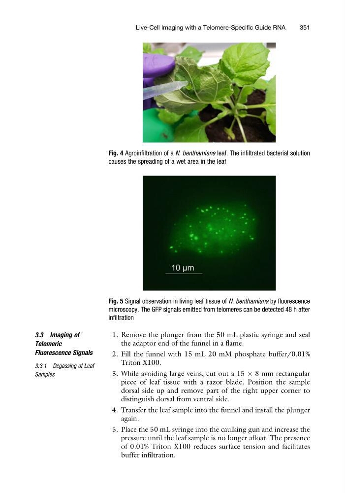
Fig. 4 Agroinfiltration of a N. benthamiana leaf. The infiltrated bacterial solution
causes the spreading of a wet area in the leaf
Fig. 5 Signal observation in living leaf tissue of N. benthamiana by fluorescence
microscopy. The GFP signals emitted from telomeres can be detected 48 h after
infiltration
Live-Cell Imaging with a Telomere-Specific Guide RNA
351
Telomeric
Fluorescence Signals
3.3.1 Degassing of Leaf
Samples
1. Remove the plunger from the 50 mL plastic syringe and seal
the adaptor end of the funnel in a flame.
2. Fill the funnel with 15 mL 20 mM phosphate buffer/0.01%
Triton X100.
3. While avoiding large veins, cut out a 15 Â 8 mm rectangular
piece of leaf tissue with a razor blade. Position the sample
dorsal side up and remove part of the right upper corner to
distinguish dorsal from ventral side.
4. Transfer the leaf sample into the funnel and install the plunger
again.
5. Place the 50 mL syringe into the caulking gun and increase the
pressure until the leaf sample is no longer afloat. The presence
of 0.01% Triton X100 reduces surface tension and facilitates
buffer infiltration.
Fig. 4 Agroinfiltration of a N. benthamiana leaf. The infiltrated bacterial solution
causes the spreading of a wet area in the leaf
Fig. 5 Signal observation in living leaf tissue of N. benthamiana by fluorescence
microscopy. The GFP signals emitted from telomeres can be detected 48 h after
infiltration
Live-Cell Imaging with a Telomere-Specific Guide RNA
351