

Before proceeding with this experiment, the genomic DNA or
cDNA encoding the proteins of interest should be cloned in translational fusion with a red fluorescent protein of choice, under the
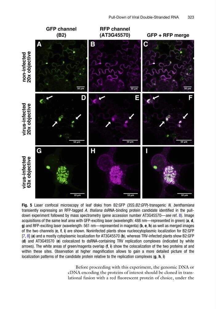
Fig. 5 Laser confocal microscopy of leaf disks from B2:GFP (35S:B2:GFP)-transgenic N. benthamiana
transiently expressing an RFP-tagged A. thaliana dsRNA-binding protein candidate identified in the pulldown experiment followed by mass spectrometry (gene accession number AT3G45570—see ref. 8). Image
acquisitions of the same leaf area with GFP-exciting laser (wavelength: 488 nm—represented in green) (a, d,
g) and RFP-exciting laser (wavelength: 561 nm—represented in magenta) (b, e, h) as well as merged images
of the two channels (c, f, i) are shown. Noninfected plants show nucleocytoplasmic localization for B2:GFP
[7, 8] (a) and a mostly cytoplasmic localization for AT3G45570 (b), whereas TRV-infected plants show B2:GFP
(d) and AT3G45570 (e) colocalized to dsRNA-containing TRV replication complexes (indicated by white
arrows). The white areas of green/magenta overlap (f, i) show the colocalization of the two proteins at and
within these sites. Observation at higher magnification allows to gain a more detailed picture of the
localization patterns of the candidate protein relative to the replication complexes (g, h, i)
Pull-Down of Viral Double-Stranded RNA
323
cDNA encoding the proteins of interest should be cloned in translational fusion with a red fluorescent protein of choice, under the
Fig. 5 Laser confocal microscopy of leaf disks from B2:GFP (35S:B2:GFP)-transgenic N. benthamiana
transiently expressing an RFP-tagged A. thaliana dsRNA-binding protein candidate identified in the pulldown experiment followed by mass spectrometry (gene accession number AT3G45570—see ref. 8). Image
acquisitions of the same leaf area with GFP-exciting laser (wavelength: 488 nm—represented in green) (a, d,
g) and RFP-exciting laser (wavelength: 561 nm—represented in magenta) (b, e, h) as well as merged images
of the two channels (c, f, i) are shown. Noninfected plants show nucleocytoplasmic localization for B2:GFP
[7, 8] (a) and a mostly cytoplasmic localization for AT3G45570 (b), whereas TRV-infected plants show B2:GFP
(d) and AT3G45570 (e) colocalized to dsRNA-containing TRV replication complexes (indicated by white
arrows). The white areas of green/magenta overlap (f, i) show the colocalization of the two proteins at and
within these sites. Observation at higher magnification allows to gain a more detailed picture of the
localization patterns of the candidate protein relative to the replication complexes (g, h, i)
Pull-Down of Viral Double-Stranded RNA
323