

by hybridization) to a PNK labeling reaction prepared according to the manufacturer’s instructions. Using filter tips, add the
32 P-labeled γATP, mix by pipetting, and incubate the mix at
37
C for 1 h. Prepare a sepharose column and remove liquid
according to the manufacturer’s instructions. Place the column
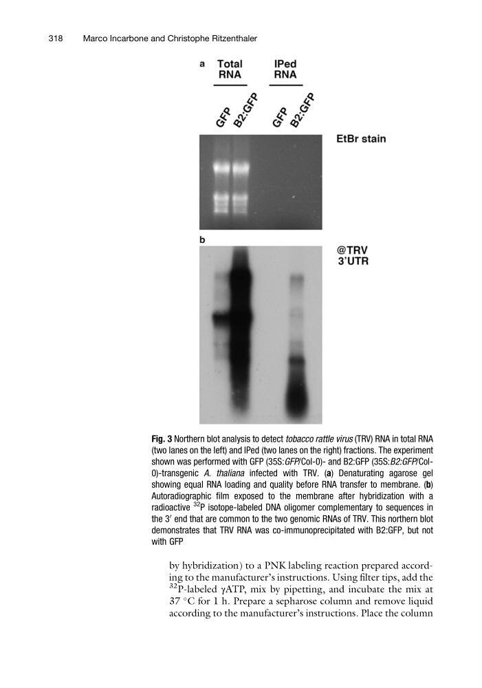
Fig. 3 Northern blot analysis to detect tobacco rattle virus (TRV) RNA in total RNA
(two lanes on the left) and IPed (two lanes on the right) fractions. The experiment
shown was performed with GFP (35S:GFP/Col-0)- and B2:GFP (35S:B2:GFP/Col0)-transgenic A. thaliana infected with TRV. (a) Denaturating agarose gel
showing equal RNA loading and quality before RNA transfer to membrane. (b)
Autoradiographic film exposed to the membrane after hybridization with a
radioactive
32 P isotope-labeled DNA oligomer complementary to sequences in
the 3
0 end that are common to the two genomic RNAs of TRV. This northern blot
demonstrates that TRV RNA was co-immunoprecipitated with B2:GFP, but not
with GFP
318
Marco Incarbone and Christophe Ritzenthaler
32 P-labeled γATP, mix by pipetting, and incubate the mix at
37
C for 1 h. Prepare a sepharose column and remove liquid
according to the manufacturer’s instructions. Place the column
Fig. 3 Northern blot analysis to detect tobacco rattle virus (TRV) RNA in total RNA
(two lanes on the left) and IPed (two lanes on the right) fractions. The experiment
shown was performed with GFP (35S:GFP/Col-0)- and B2:GFP (35S:B2:GFP/Col0)-transgenic A. thaliana infected with TRV. (a) Denaturating agarose gel
showing equal RNA loading and quality before RNA transfer to membrane. (b)
Autoradiographic film exposed to the membrane after hybridization with a
radioactive
32 P isotope-labeled DNA oligomer complementary to sequences in
the 3
0 end that are common to the two genomic RNAs of TRV. This northern blot
demonstrates that TRV RNA was co-immunoprecipitated with B2:GFP, but not
with GFP
318
Marco Incarbone and Christophe Ritzenthaler