

6. In a sterile hood, pipette 1 mL hot liquid MEA medium
(at around 60
C) onto each sterile microscope slide and
allow the MEA to be evenly and fully distributed onto each
slide (see Note 6).
7. Wait about 5–10 min for the MEA medium to solidify.
8. Add 4 μL of 100 ng/μL fluorescent sRNAs or dsRNAs to 4 μL
of the 1 Â 10
5 spores/mL suspension. As a control, add 4 μL
of sterile water and 4 μL of properly diluted fluorescein (see
Note 7) to 4 μL of the 1 Â 10
5 spores/mL suspension (see
Note 8).
9. Directly apply these solutions to the MEA microscope slides.
10. Incubate the microscope slides in a sterile petri dish covered
with aluminum foil to prevent direct exposure to the light at
room temperature for 12 h (see Note 9).
11. Treat mycelium with 20 μL 75 U micrococcal nuclease at 37
C
for 30 min to degrade any fluorescent dsRNA and sRNAs
outside of the fungal mycelium.
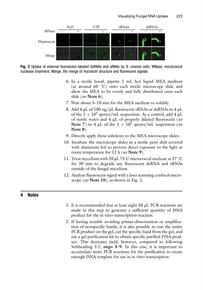
12. Analyze fluorescent signal with a laser scanning confocal microscope (see Note 10), as shown in Fig. 2.
4 Notes
1. It is recommended that at least eight 50 μL PCR reactions are
made in this step to generate a sufficient quantity of DNA
product for the in vitro transcription reaction.
2. If having trouble avoiding primer-dimerization or amplification of nonspecific bands, it is also possible to run the entire
PCR product on the gel, cut the specific band from the gel, and
use a gel purification kit to obtain specific purified DNA product. This decreases yield, however, compared to following
Subheading 3.1, steps 3–9. In this case, it is important to
accumulate more PCR reactions for the purification to create
enough DNA template for use in in vitro transcription.
Fluorescein
MNase
-
+
dsRNAs
-
+
sRNAs
-
+
H 2 O
Merge
-
+
UTP
Fig. 2 Uptake of external fluorescein-labeled dsRNAs and sRNAs by B. cinerea cells. MNase, micrococcal
nuclease treatment. Merge, the merge of mycelium structure and fluorescein signals
Visualizing Fungal RNA Uptake
223
(at around 60
C) onto each sterile microscope slide and
allow the MEA to be evenly and fully distributed onto each
slide (see Note 6).
7. Wait about 5–10 min for the MEA medium to solidify.
8. Add 4 μL of 100 ng/μL fluorescent sRNAs or dsRNAs to 4 μL
of the 1 Â 10
5 spores/mL suspension. As a control, add 4 μL
of sterile water and 4 μL of properly diluted fluorescein (see
Note 7) to 4 μL of the 1 Â 10
5 spores/mL suspension (see
Note 8).
9. Directly apply these solutions to the MEA microscope slides.
10. Incubate the microscope slides in a sterile petri dish covered
with aluminum foil to prevent direct exposure to the light at
room temperature for 12 h (see Note 9).
11. Treat mycelium with 20 μL 75 U micrococcal nuclease at 37
C
for 30 min to degrade any fluorescent dsRNA and sRNAs
outside of the fungal mycelium.
12. Analyze fluorescent signal with a laser scanning confocal microscope (see Note 10), as shown in Fig. 2.
4 Notes
1. It is recommended that at least eight 50 μL PCR reactions are
made in this step to generate a sufficient quantity of DNA
product for the in vitro transcription reaction.
2. If having trouble avoiding primer-dimerization or amplification of nonspecific bands, it is also possible to run the entire
PCR product on the gel, cut the specific band from the gel, and
use a gel purification kit to obtain specific purified DNA product. This decreases yield, however, compared to following
Subheading 3.1, steps 3–9. In this case, it is important to
accumulate more PCR reactions for the purification to create
enough DNA template for use in in vitro transcription.
Fluorescein
MNase
-
+
dsRNAs
-
+
sRNAs
-
+
H 2 O
Merge
-
+
UTP
Fig. 2 Uptake of external fluorescein-labeled dsRNAs and sRNAs by B. cinerea cells. MNase, micrococcal
nuclease treatment. Merge, the merge of mycelium structure and fluorescein signals
Visualizing Fungal RNA Uptake
223