

4. Entry clones can be obtained either by traditional restriction
endonuclease/ligase methods, through a BP™ reaction in
which the gene of interest is flanked by recombination sites
by PCR and then recombined into a donor vector
(pDONR™), or by TOPO™ cloning. The three alternatives
offer directional cloning, but special considerations are needed
to design primers and ensure the desired cloning result. Please
refer
to
http://tools.thermofisher.com/content/sfs/
manuals/gatewayman.pdf for instructions for the design of
primers appropriate for BP cloning into pDONR™ vectors,
and to https://assets.thermofisher.com/TFS-Assets/LSG/
manuals/pentr_dtopo_man.pdf for instructions for the design
of primers suitable for TOPO
® cloning into pENTR™ vectors.
5. For co-expression experiments, the bacterial cultures resuspended in sterile distilled water are mixed before infiltration.
To obtain the desired expression level, the concentration of
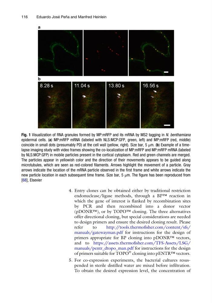
Fig. 1 Visualization of RNA granules formed by MP:mRFP and its mRNA by MS2 tagging in N. benthamiana
epidermal cells. (a) MP:mRFP mRNA (labeled with NLS:MCP:GFP, green, left) and MP:mRFP (red, middle)
coincide in small dots (presumably PD) at the cell wall (yellow, right). Size bar, 5 μm. (b) Example of a timelapse imaging study with video frames showing the co-localization of MP:mRFP and MP:mRFP mRNA (labeled
by NLS:MCP:GFP) in mobile particles present in the cortical cytoplasm. Red and green channels are merged.
The particles appear in yellowish color and the direction of their movements appears to be guided along
microtubules, which are seen as red-colored filaments. Arrows highlight the movement of a particle. Gray
arrows indicate the location of the mRNA particle observed in the first frame and white arrows indicate the
new particle location in each subsequent time frame. Size bar, 5 μm. The figure has been reproduced from
[68], Elsevier
116
Eduardo Jose ´ Pen ˜ a and Manfred Heinlein
endonuclease/ligase methods, through a BP™ reaction in
which the gene of interest is flanked by recombination sites
by PCR and then recombined into a donor vector
(pDONR™), or by TOPO™ cloning. The three alternatives
offer directional cloning, but special considerations are needed
to design primers and ensure the desired cloning result. Please
refer
to
http://tools.thermofisher.com/content/sfs/
manuals/gatewayman.pdf for instructions for the design of
primers appropriate for BP cloning into pDONR™ vectors,
and to https://assets.thermofisher.com/TFS-Assets/LSG/
manuals/pentr_dtopo_man.pdf for instructions for the design
of primers suitable for TOPO
® cloning into pENTR™ vectors.
5. For co-expression experiments, the bacterial cultures resuspended in sterile distilled water are mixed before infiltration.
To obtain the desired expression level, the concentration of
Fig. 1 Visualization of RNA granules formed by MP:mRFP and its mRNA by MS2 tagging in N. benthamiana
epidermal cells. (a) MP:mRFP mRNA (labeled with NLS:MCP:GFP, green, left) and MP:mRFP (red, middle)
coincide in small dots (presumably PD) at the cell wall (yellow, right). Size bar, 5 μm. (b) Example of a timelapse imaging study with video frames showing the co-localization of MP:mRFP and MP:mRFP mRNA (labeled
by NLS:MCP:GFP) in mobile particles present in the cortical cytoplasm. Red and green channels are merged.
The particles appear in yellowish color and the direction of their movements appears to be guided along
microtubules, which are seen as red-colored filaments. Arrows highlight the movement of a particle. Gray
arrows indicate the location of the mRNA particle observed in the first frame and white arrows indicate the
new particle location in each subsequent time frame. Size bar, 5 μm. The figure has been reproduced from
[68], Elsevier
116
Eduardo Jose ´ Pen ˜ a and Manfred Heinlein