

6.2.4 Probing of Intracellular Environment by SERS
Imaging
Although Raman microscopy has been widely employed for imaging of cells, it is
limited because of a weak signal in comparison with the background noise or
autofluorescence. Therefore, as data collection process is time-consuming, reliable
statistical analysis of weakly scattering biosamples is hindered [118]. This obstacle
can be overcome by using NPs as a SERS agent to enhance Raman scattering
(Sect. 6.1.4) and to gather information on specific cellular processes as well as type
of molecules and changes in the intracellular environment. Intracellular SERS
detection is usually accomplished by two strategies, i.e., label (indirect) and
label-free (direct) methods. In the latter, bare (‘naked’) Au or Ag nanoparticles or
nanoaggregates are used to interact with cellular biomolecules [119, 120]. An
effective and successful utilization of NPs for in vitro SERS analysis requires
understanding of pathways and mechanisms of their uptake and distribution.
Endocytosis and passive diffusion are known to be a major pathway for AuNPs
[121, 122]. Metallic nanoparticles after uptake by cells are mostly localized in
endosomes; therefore, the SERS intracellular spectra represent localization of these
structures, their composition, and local environment [47, 122]. Kneipp et al. [123]
have showed that a label-free method of probing living macrophages and
endothelial cells differentiates cell lines and characterizes changes in cellular
environments by detection of physiologically relevant molecules. This method has
been successfully employed in elucidating biomolecular dynamics of stress
response upon UV-C irradiation at a cellular level in healthy and cancerous cells.
Functionalization of Au nanocubes with nuclear localization signal peptide and
cell-penetrating peptide enabled observing physiochemical damages of cytosolic
proteins containing sulfur and aromatic amino acids by changes in their secondary
structures showing that SERS was a powerful approach to understand mechanisms
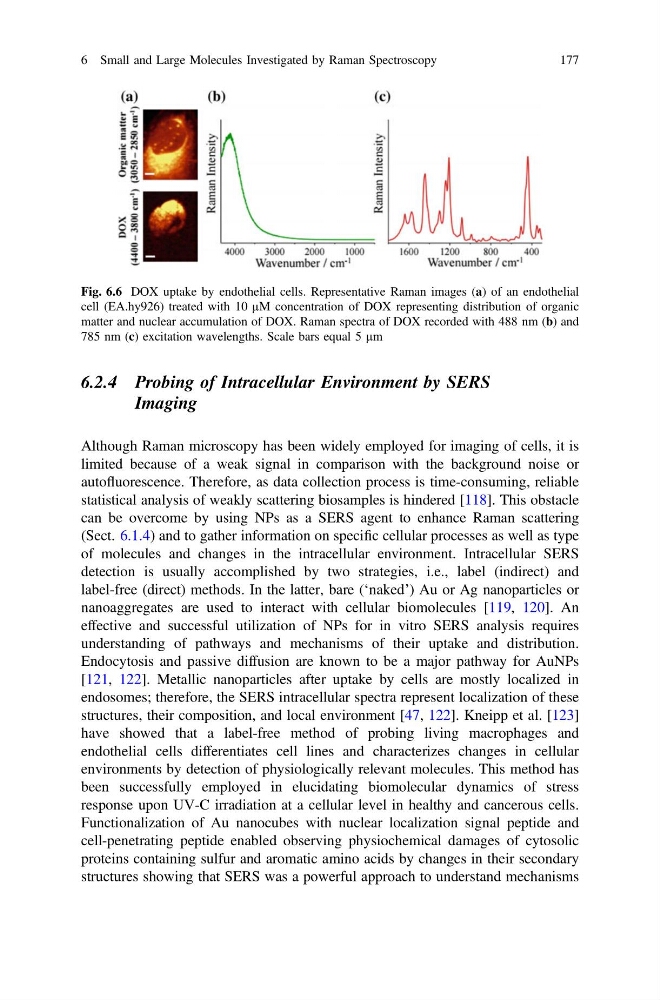
Fig. 6.6 DOX uptake by endothelial cells. Representative Raman images (a) of an endothelial
cell (EA.hy926) treated with 10 µM concentration of DOX representing distribution of organic
matter and nuclear accumulation of DOX. Raman spectra of DOX recorded with 488 nm (b) and
785 nm (c) excitation wavelengths. Scale bars equal 5 lm
6 Small and Large Molecules Investigated by Raman Spectroscopy
177
Imaging
Although Raman microscopy has been widely employed for imaging of cells, it is
limited because of a weak signal in comparison with the background noise or
autofluorescence. Therefore, as data collection process is time-consuming, reliable
statistical analysis of weakly scattering biosamples is hindered [118]. This obstacle
can be overcome by using NPs as a SERS agent to enhance Raman scattering
(Sect. 6.1.4) and to gather information on specific cellular processes as well as type
of molecules and changes in the intracellular environment. Intracellular SERS
detection is usually accomplished by two strategies, i.e., label (indirect) and
label-free (direct) methods. In the latter, bare (‘naked’) Au or Ag nanoparticles or
nanoaggregates are used to interact with cellular biomolecules [119, 120]. An
effective and successful utilization of NPs for in vitro SERS analysis requires
understanding of pathways and mechanisms of their uptake and distribution.
Endocytosis and passive diffusion are known to be a major pathway for AuNPs
[121, 122]. Metallic nanoparticles after uptake by cells are mostly localized in
endosomes; therefore, the SERS intracellular spectra represent localization of these
structures, their composition, and local environment [47, 122]. Kneipp et al. [123]
have showed that a label-free method of probing living macrophages and
endothelial cells differentiates cell lines and characterizes changes in cellular
environments by detection of physiologically relevant molecules. This method has
been successfully employed in elucidating biomolecular dynamics of stress
response upon UV-C irradiation at a cellular level in healthy and cancerous cells.
Functionalization of Au nanocubes with nuclear localization signal peptide and
cell-penetrating peptide enabled observing physiochemical damages of cytosolic
proteins containing sulfur and aromatic amino acids by changes in their secondary
structures showing that SERS was a powerful approach to understand mechanisms
Fig. 6.6 DOX uptake by endothelial cells. Representative Raman images (a) of an endothelial
cell (EA.hy926) treated with 10 µM concentration of DOX representing distribution of organic
matter and nuclear accumulation of DOX. Raman spectra of DOX recorded with 488 nm (b) and
785 nm (c) excitation wavelengths. Scale bars equal 5 lm
6 Small and Large Molecules Investigated by Raman Spectroscopy
177