

scientific innovations, such as endogenous fluorescent proteins [1] to be applicable
to MPE microscopy. Likewise, exogenous markers like organic dyes have been
synthesized for MPE microscopy, but their use remains limited by their relatively
low two-photon excited fluorescence cross sections in aqueous environments and
their rapid photo-bleaching [2]. In addition, other probes have been proposed
including quantum and polymer nanodots with broad absorption and discrete,
tunable emission wavelengths withstanding photo-bleaching [3]. Preliminary
studies have clearly demonstrated that noble metal nanoclusters (NCs), nanomaterials made of few to hundred gold or silver atoms protected by ligand molecules
exhibiting molecular-like properties [4], could also provide a valuable route in the
nonlinear optical regime and in particular MPE microscopy [5].
Multi-photon optics stems from the nonlinear light–matter interaction and can be
described by a medium polarization P, induced by an intense optical electric field
E as:
P ¼ v
ð1Þ E þ v
ð2Þ EE þ v
ð3Þ EEE þ . . .
ð5:1Þ
where v
ð1Þ is the linear susceptibility tensor representing effects such as linear
absorption and refraction, v
ð2Þ is the second-order nonlinear optical susceptibility,
v
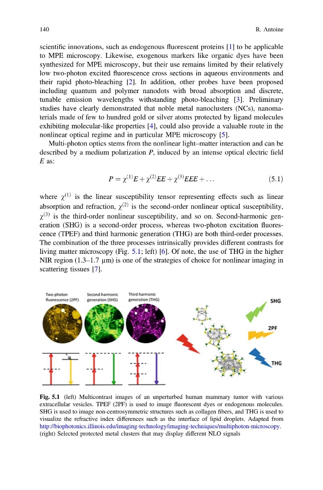
ð3Þ is the third-order nonlinear susceptibility, and so on. Second-harmonic generation (SHG) is a second-order process, whereas two-photon excitation fluorescence (TPEF) and third harmonic generation (THG) are both third-order processes.
The combination of the three processes intrinsically provides different contrasts for
living matter microscopy (Fig. 5.1; left) [6]. Of note, the use of THG in the higher
NIR region (1.3–1.7 µm) is one of the strategies of choice for nonlinear imaging in
scattering tissues [7].
Fig. 5.1 (left) Multicontrast images of an unperturbed human mammary tumor with various
extracellular vesicles. TPEF (2PF) is used to image fluorescent dyes or endogenous molecules.
SHG is used to image non-centrosymmetric structures such as collagen fibers, and THG is used to
visualize the refractive index differences such as the interface of lipid droplets. Adapted from
http://biophotonics.illinois.edu/imaging-technology/imaging-techniques/multiphoton-microscopy.
(right) Selected protected metal clusters that may display different NLO signals
140
R. Antoine
to MPE microscopy. Likewise, exogenous markers like organic dyes have been
synthesized for MPE microscopy, but their use remains limited by their relatively
low two-photon excited fluorescence cross sections in aqueous environments and
their rapid photo-bleaching [2]. In addition, other probes have been proposed
including quantum and polymer nanodots with broad absorption and discrete,
tunable emission wavelengths withstanding photo-bleaching [3]. Preliminary
studies have clearly demonstrated that noble metal nanoclusters (NCs), nanomaterials made of few to hundred gold or silver atoms protected by ligand molecules
exhibiting molecular-like properties [4], could also provide a valuable route in the
nonlinear optical regime and in particular MPE microscopy [5].
Multi-photon optics stems from the nonlinear light–matter interaction and can be
described by a medium polarization P, induced by an intense optical electric field
E as:
P ¼ v
ð1Þ E þ v
ð2Þ EE þ v
ð3Þ EEE þ . . .
ð5:1Þ
where v
ð1Þ is the linear susceptibility tensor representing effects such as linear
absorption and refraction, v
ð2Þ is the second-order nonlinear optical susceptibility,
v
ð3Þ is the third-order nonlinear susceptibility, and so on. Second-harmonic generation (SHG) is a second-order process, whereas two-photon excitation fluorescence (TPEF) and third harmonic generation (THG) are both third-order processes.
The combination of the three processes intrinsically provides different contrasts for
living matter microscopy (Fig. 5.1; left) [6]. Of note, the use of THG in the higher
NIR region (1.3–1.7 µm) is one of the strategies of choice for nonlinear imaging in
scattering tissues [7].
Fig. 5.1 (left) Multicontrast images of an unperturbed human mammary tumor with various
extracellular vesicles. TPEF (2PF) is used to image fluorescent dyes or endogenous molecules.
SHG is used to image non-centrosymmetric structures such as collagen fibers, and THG is used to
visualize the refractive index differences such as the interface of lipid droplets. Adapted from
http://biophotonics.illinois.edu/imaging-technology/imaging-techniques/multiphoton-microscopy.
(right) Selected protected metal clusters that may display different NLO signals
140
R. Antoine