

5. At the end of the incubation period, vortex vigorously again for
10 s.
6. Set the amplitude and the time on the sonicator to 35% intensity and 1 min, respectively. Be sure that the sonicator probe has
been cleaned first with water and then 70% ethanol twice
through and wiped clean with paper cloth.
7. While the sample is kept on ice, sonicate each sample for 1 min
and place it on ice for 1 min. Repeat the procedure four times.
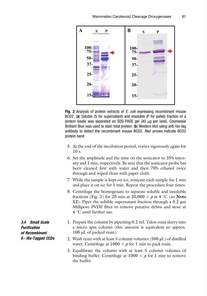
8. Centrifuge the homogenate to separate soluble and insoluble
fractions (Fig. 2) for 20 min at 20,000 Â g at 4
C (see Note
12). Pipet the soluble supernatant fraction through a 0.2 μm
Millipore PVDF filter to remove putative debris and store at
4
C until further use.
3.4 Small Scale
Purification
of Recombinant
6ÂHis-Tagged CCDs
1. Prepare the column by pipetting 0.2 mL Talon resin slurry into
a micro spin column (this amount is equivalent to approx.
100 μL of packed resin).
2. Wash resin with at least 5 column volumes (500 μL) of distilled
water. Centrifuge at 1000 Â g for 1 min to pack resin.
3. Equilibrate the column with at least 5 column volumes of
binding buffer. Centrifuge at 1000 Â g for 1 min to remove
the buffer.
Fig. 2 Analysis of protein extracts of E. coli expressing recombinant mouse
BCO2. (a) Soluble (S for supernatant) and insoluble (P for pellet) fraction of a
protein lysate was separated on SDS-PAGE gel (40 μg per lane). Coomassie
Brilliant Blue was used to stain total protein. (b) Western blot using anti-his-tag
antibody to detect the recombinant mouse BCO2. Red arrows indicate BCO2
protein band
Mammalian Carotenoid Cleavage Dioxygenases
81
10 s.
6. Set the amplitude and the time on the sonicator to 35% intensity and 1 min, respectively. Be sure that the sonicator probe has
been cleaned first with water and then 70% ethanol twice
through and wiped clean with paper cloth.
7. While the sample is kept on ice, sonicate each sample for 1 min
and place it on ice for 1 min. Repeat the procedure four times.
8. Centrifuge the homogenate to separate soluble and insoluble
fractions (Fig. 2) for 20 min at 20,000 Â g at 4
C (see Note
12). Pipet the soluble supernatant fraction through a 0.2 μm
Millipore PVDF filter to remove putative debris and store at
4
C until further use.
3.4 Small Scale
Purification
of Recombinant
6ÂHis-Tagged CCDs
1. Prepare the column by pipetting 0.2 mL Talon resin slurry into
a micro spin column (this amount is equivalent to approx.
100 μL of packed resin).
2. Wash resin with at least 5 column volumes (500 μL) of distilled
water. Centrifuge at 1000 Â g for 1 min to pack resin.
3. Equilibrate the column with at least 5 column volumes of
binding buffer. Centrifuge at 1000 Â g for 1 min to remove
the buffer.
Fig. 2 Analysis of protein extracts of E. coli expressing recombinant mouse
BCO2. (a) Soluble (S for supernatant) and insoluble (P for pellet) fraction of a
protein lysate was separated on SDS-PAGE gel (40 μg per lane). Coomassie
Brilliant Blue was used to stain total protein. (b) Western blot using anti-his-tag
antibody to detect the recombinant mouse BCO2. Red arrows indicate BCO2
protein band
Mammalian Carotenoid Cleavage Dioxygenases
81