

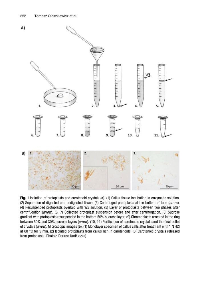
Fig. 1 Isolation of protoplasts and carotenoid crystals (a). (1) Callus tissue incubation in enzymatic solution.
(2) Separation of digested and undigested tissue. (3) Centrifuged protoplasts at the bottom of tube (arrow).
(4) Resuspended protoplasts overlaid with W5 solution. (5) Layer of protoplasts between two phases after
centrifugation (arrow). (6, 7) Collected protoplast suspension before and after centrifugation. (8) Sucrose
gradient with protoplasts resuspended in the bottom 50% sucrose layer. (9) Chromoplasts arrested in the ring
between 50% and 30% sucrose layers (arrow). (10, 11) Purification of carotenoid crystals and the final pellet
of crystals (arrow). Microscopic images (b). (1) Monolayer specimen of callus cells after treatment with 1 N HCl
at 60
C for 5 min. (2) Isolated protoplasts from callus rich in carotenoids. (3) Carotenoid crystals released
from protoplasts (Photos: Dariusz Kadłuczka)
252
Tomasz Oleszkiewicz et al.
(2) Separation of digested and undigested tissue. (3) Centrifuged protoplasts at the bottom of tube (arrow).
(4) Resuspended protoplasts overlaid with W5 solution. (5) Layer of protoplasts between two phases after
centrifugation (arrow). (6, 7) Collected protoplast suspension before and after centrifugation. (8) Sucrose
gradient with protoplasts resuspended in the bottom 50% sucrose layer. (9) Chromoplasts arrested in the ring
between 50% and 30% sucrose layers (arrow). (10, 11) Purification of carotenoid crystals and the final pellet
of crystals (arrow). Microscopic images (b). (1) Monolayer specimen of callus cells after treatment with 1 N HCl
at 60
C for 5 min. (2) Isolated protoplasts from callus rich in carotenoids. (3) Carotenoid crystals released
from protoplasts (Photos: Dariusz Kadłuczka)
252
Tomasz Oleszkiewicz et al.