

79
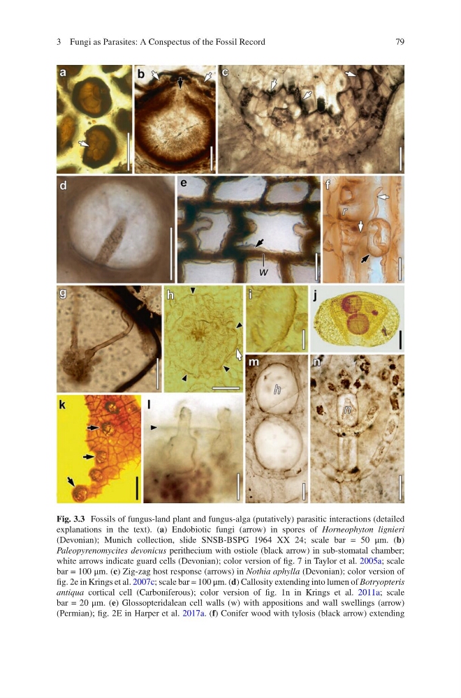
Fig. 3.3 Fossils of fungus-land plant and fungus-alga (putatively) parasitic interactions (detailed
explanations in the text). (a) Endobiotic fungi (arrow) in spores of Horneophyton lignieri
(Devonian); Munich collection, slide SNSB-BSPG 1964 XX 24; scale bar = 50 μm. (b)
Paleopyrenomycites devonicus perithecium with ostiole (black arrow) in sub-stomatal chamber;
white arrows indicate guard cells (Devonian); color version of fig. 7 in Taylor et al. 2005a; scale
bar = 100 μm. (c) Zig-zag host response (arrows) in Nothia aphylla (Devonian); color version of
fig. 2e in Krings et al. 2007c; scale bar = 100 μm. (d) Callosity extending into lumen of Botryopteris
antiqua cortical cell (Carboniferous); color version of fig. 1n in Krings et al. 2011a; scale
bar = 20 μm. (e) Glossopteridalean cell walls (w) with appositions and wall swellings (arrow)
(Permian); fig. 2E in Harper et al. 2017a. (f) Conifer wood with tylosis (black arrow) extending
3 Fungi as Parasites: A Conspectus of the Fossil Record
Fig. 3.3 Fossils of fungus-land plant and fungus-alga (putatively) parasitic interactions (detailed
explanations in the text). (a) Endobiotic fungi (arrow) in spores of Horneophyton lignieri
(Devonian); Munich collection, slide SNSB-BSPG 1964 XX 24; scale bar = 50 μm. (b)
Paleopyrenomycites devonicus perithecium with ostiole (black arrow) in sub-stomatal chamber;
white arrows indicate guard cells (Devonian); color version of fig. 7 in Taylor et al. 2005a; scale
bar = 100 μm. (c) Zig-zag host response (arrows) in Nothia aphylla (Devonian); color version of
fig. 2e in Krings et al. 2007c; scale bar = 100 μm. (d) Callosity extending into lumen of Botryopteris
antiqua cortical cell (Carboniferous); color version of fig. 1n in Krings et al. 2011a; scale
bar = 20 μm. (e) Glossopteridalean cell walls (w) with appositions and wall swellings (arrow)
(Permian); fig. 2E in Harper et al. 2017a. (f) Conifer wood with tylosis (black arrow) extending
3 Fungi as Parasites: A Conspectus of the Fossil Record