

44
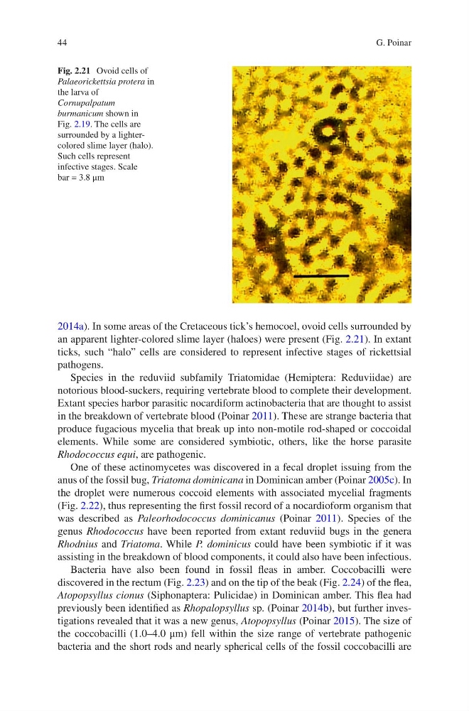
2014a). In some areas of the Cretaceous tick’s hemocoel, ovoid cells surrounded by
an apparent lighter-colored slime layer (haloes) were present (Fig. 2.21). In extant
ticks, such “halo” cells are considered to represent infective stages of rickettsial
pathogens.
Species in the reduviid subfamily Triatomidae (Hemiptera: Reduviidae) are
notorious blood-suckers, requiring vertebrate blood to complete their development.
Extant species harbor parasitic nocardiform actinobacteria that are thought to assist
in the breakdown of vertebrate blood (Poinar 2011). These are strange bacteria that
produce fugacious mycelia that break up into non-motile rod-shaped or coccoidal
elements. While some are considered symbiotic, others, like the horse parasite
Rhodococcus equi, are pathogenic.
One of these actinomycetes was discovered in a fecal droplet issuing from the
anus of the fossil bug, Triatoma dominicana in Dominican amber (Poinar 2005c). In
the droplet were numerous coccoid elements with associated mycelial fragments
(Fig. 2.22), thus representing the first fossil record of a nocardioform organism that
was described as Paleorhodococcus dominicanus (Poinar 2011). Species of the
genus Rhodococcus have been reported from extant reduviid bugs in the genera
Rhodnius and Triatoma. While P. dominicus could have been symbiotic if it was
assisting in the breakdown of blood components, it could also have been infectious.
Bacteria have also been found in fossil fleas in amber. Coccobacilli were
discovered in the rectum (Fig. 2.23) and on the tip of the beak (Fig. 2.24) of the flea,
Atopopsyllus cionus (Siphonaptera: Pulicidae) in Dominican amber. This flea had
previously been identified as Rhopalopsyllus sp. (Poinar 2014b), but further investigations revealed that it was a new genus, Atopopsyllus (Poinar 2015). The size of
the coccobacilli (1.0–4.0 μm) fell within the size range of vertebrate pathogenic
bacteria and the short rods and nearly spherical cells of the fossil coccobacilli are
Fig. 2.21 Ovoid cells of
Palaeorickettsia protera in
the larva of
Cornupalpatum
burmanicum shown in
Fig. 2.19. The cells are
surrounded by a lightercolored slime layer (halo).
Such cells represent
infective stages. Scale
bar = 3.8 μm
G. Poinar
2014a). In some areas of the Cretaceous tick’s hemocoel, ovoid cells surrounded by
an apparent lighter-colored slime layer (haloes) were present (Fig. 2.21). In extant
ticks, such “halo” cells are considered to represent infective stages of rickettsial
pathogens.
Species in the reduviid subfamily Triatomidae (Hemiptera: Reduviidae) are
notorious blood-suckers, requiring vertebrate blood to complete their development.
Extant species harbor parasitic nocardiform actinobacteria that are thought to assist
in the breakdown of vertebrate blood (Poinar 2011). These are strange bacteria that
produce fugacious mycelia that break up into non-motile rod-shaped or coccoidal
elements. While some are considered symbiotic, others, like the horse parasite
Rhodococcus equi, are pathogenic.
One of these actinomycetes was discovered in a fecal droplet issuing from the
anus of the fossil bug, Triatoma dominicana in Dominican amber (Poinar 2005c). In
the droplet were numerous coccoid elements with associated mycelial fragments
(Fig. 2.22), thus representing the first fossil record of a nocardioform organism that
was described as Paleorhodococcus dominicanus (Poinar 2011). Species of the
genus Rhodococcus have been reported from extant reduviid bugs in the genera
Rhodnius and Triatoma. While P. dominicus could have been symbiotic if it was
assisting in the breakdown of blood components, it could also have been infectious.
Bacteria have also been found in fossil fleas in amber. Coccobacilli were
discovered in the rectum (Fig. 2.23) and on the tip of the beak (Fig. 2.24) of the flea,
Atopopsyllus cionus (Siphonaptera: Pulicidae) in Dominican amber. This flea had
previously been identified as Rhopalopsyllus sp. (Poinar 2014b), but further investigations revealed that it was a new genus, Atopopsyllus (Poinar 2015). The size of
the coccobacilli (1.0–4.0 μm) fell within the size range of vertebrate pathogenic
bacteria and the short rods and nearly spherical cells of the fossil coccobacilli are
Fig. 2.21 Ovoid cells of
Palaeorickettsia protera in
the larva of
Cornupalpatum
burmanicum shown in
Fig. 2.19. The cells are
surrounded by a lightercolored slime layer (halo).
Such cells represent
infective stages. Scale
bar = 3.8 μm
G. Poinar