

277
functional unit named presoma (also praesoma). Farther posterior follows the trunk
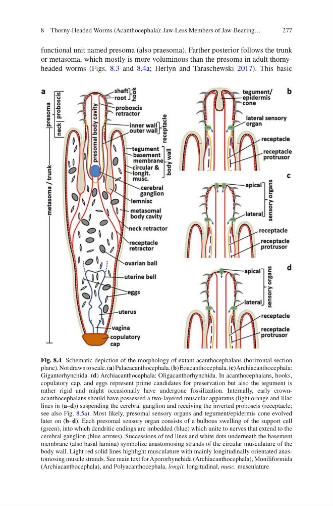
or metasoma, which mostly is more voluminous than the presoma in adult thornyheaded worms (Figs. 8.3 and 8.4a; Herlyn and Taraschewski 2017). This basic
Fig. 8.4 Schematic depiction of the morphology of extant acanthocephalans (horizontal section
plane). Not drawn to scale. (a) Palaeacanthocephala. (b) Eoacanthocephala. (c) Archiacanthocephala:
Gigantorhynchida. (d) Archiacanthocephala: Oligacanthorhynchida. In acanthocephalans, hooks,
copulatory cap, and eggs represent prime candidates for preservation but also the tegument is
rather rigid and might occasionally have undergone fossilization. Internally, early crownacanthocephalans should have possessed a two-layered muscular apparatus (light orange and lilac
lines in (a–d)) suspending the cerebral ganglion and receiving the inverted proboscis (receptacle;
see also Fig. 8.5a). Most likely, presomal sensory organs and tegument/epidermis cone evolved
later on (b–d). Each presomal sensory organ consists of a bulbous swelling of the support cell
(green), into which dendritic endings are imbedded (blue) which unite to nerves that extend to the
cerebral ganglion (blue arrows). Successions of red lines and white dots underneath the basement
membrane (also basal lamina) symbolize anastomosing strands of the circular musculature of the
body wall. Light red solid lines highlight musculature with mainly longitudinally orientated anastomosing muscle strands. See main text for Apororhynchida (Archiacanthocephala), Moniliformida
(Archiacanthocephala), and Polyacanthocephala. longit. longitudinal, musc. musculature
8 Thorny-Headed Worms (Acanthocephala): Jaw-Less Members of Jaw-Bearing…
functional unit named presoma (also praesoma). Farther posterior follows the trunk
or metasoma, which mostly is more voluminous than the presoma in adult thornyheaded worms (Figs. 8.3 and 8.4a; Herlyn and Taraschewski 2017). This basic
Fig. 8.4 Schematic depiction of the morphology of extant acanthocephalans (horizontal section
plane). Not drawn to scale. (a) Palaeacanthocephala. (b) Eoacanthocephala. (c) Archiacanthocephala:
Gigantorhynchida. (d) Archiacanthocephala: Oligacanthorhynchida. In acanthocephalans, hooks,
copulatory cap, and eggs represent prime candidates for preservation but also the tegument is
rather rigid and might occasionally have undergone fossilization. Internally, early crownacanthocephalans should have possessed a two-layered muscular apparatus (light orange and lilac
lines in (a–d)) suspending the cerebral ganglion and receiving the inverted proboscis (receptacle;
see also Fig. 8.5a). Most likely, presomal sensory organs and tegument/epidermis cone evolved
later on (b–d). Each presomal sensory organ consists of a bulbous swelling of the support cell
(green), into which dendritic endings are imbedded (blue) which unite to nerves that extend to the
cerebral ganglion (blue arrows). Successions of red lines and white dots underneath the basement
membrane (also basal lamina) symbolize anastomosing strands of the circular musculature of the
body wall. Light red solid lines highlight musculature with mainly longitudinally orientated anastomosing muscle strands. See main text for Apororhynchida (Archiacanthocephala), Moniliformida
(Archiacanthocephala), and Polyacanthocephala. longit. longitudinal, musc. musculature
8 Thorny-Headed Worms (Acanthocephala): Jaw-Less Members of Jaw-Bearing…