

183
et al. 2005; Rogers-Lowery and Dimock 2006; Barnhart et al. 2008). Today, glochidia
are completely enclosed by single or multiple layers of host cells within 2–36 h (Lefevre
and Curtis 1910b; Nezlin et al. 1994; Jansen et al. 2001; Araujo et al. 2002; Howerth and
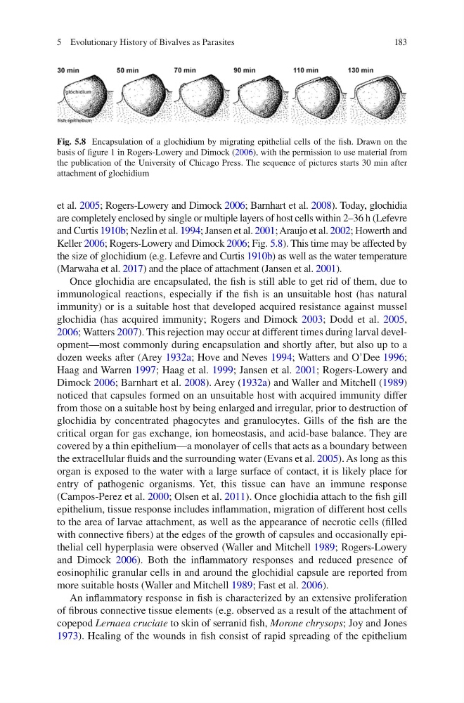
Keller 2006; Rogers-Lowery and Dimock 2006; Fig. 5.8). This time may be affected by
the size of glochidium (e.g. Lefevre and Curtis 1910b) as well as the water temperature
(Marwaha et al. 2017) and the place of attachment (Jansen et al. 2001).
Once glochidia are encapsulated, the fish is still able to get rid of them, due to
immunological reactions, especially if the fish is an unsuitable host (has natural
immunity) or is a suitable host that developed acquired resistance against mussel
glochidia (has acquired immunity; Rogers and Dimock 2003; Dodd et al. 2005,
2006; Watters 2007). This rejection may occur at different times during larval development—most commonly during encapsulation and shortly after, but also up to a
dozen weeks after (Arey 1932a; Hove and Neves 1994; Watters and O’Dee 1996;
Haag and Warren 1997; Haag et al. 1999; Jansen et al. 2001; Rogers-Lowery and
Dimock 2006; Barnhart et al. 2008). Arey (1932a) and Waller and Mitchell (1989)
noticed that capsules formed on an unsuitable host with acquired immunity differ
from those on a suitable host by being enlarged and irregular, prior to destruction of
glochidia by concentrated phagocytes and granulocytes. Gills of the fish are the
critical organ for gas exchange, ion homeostasis, and acid-base balance. They are
covered by a thin epithelium—a monolayer of cells that acts as a boundary between
the extracellular fluids and the surrounding water (Evans et al. 2005). As long as this
organ is exposed to the water with a large surface of contact, it is likely place for
entry of pathogenic organisms. Yet, this tissue can have an immune response
(Campos-Perez et al. 2000; Olsen et al. 2011). Once glochidia attach to the fish gill
epithelium, tissue response includes inflammation, migration of different host cells
to the area of larvae attachment, as well as the appearance of necrotic cells (filled
with connective fibers) at the edges of the growth of capsules and occasionally epithelial cell hyperplasia were observed (Waller and Mitchell 1989; Rogers-Lowery
and Dimock 2006). Both the inflammatory responses and reduced presence of
eosinophilic granular cells in and around the glochidial capsule are reported from
more suitable hosts (Waller and Mitchell 1989; Fast et al. 2006).
An inflammatory response in fish is characterized by an extensive proliferation
of fibrous connective tissue elements (e.g. observed as a result of the attachment of
copepod Lernaea cruciate to skin of serranid fish, Morone chrysops; Joy and Jones
1973). Healing of the wounds in fish consist of rapid spreading of the epithelium
Fig. 5.8 Encapsulation of a glochidium by migrating epithelial cells of the fish. Drawn on the
basis of figure 1 in Rogers-Lowery and Dimock (2006), with the permission to use material from
the publication of the University of Chicago Press. The sequence of pictures starts 30 min after
attachment of glochidium
5 Evolutionary History of Bivalves as Parasites
et al. 2005; Rogers-Lowery and Dimock 2006; Barnhart et al. 2008). Today, glochidia
are completely enclosed by single or multiple layers of host cells within 2–36 h (Lefevre
and Curtis 1910b; Nezlin et al. 1994; Jansen et al. 2001; Araujo et al. 2002; Howerth and
Keller 2006; Rogers-Lowery and Dimock 2006; Fig. 5.8). This time may be affected by
the size of glochidium (e.g. Lefevre and Curtis 1910b) as well as the water temperature
(Marwaha et al. 2017) and the place of attachment (Jansen et al. 2001).
Once glochidia are encapsulated, the fish is still able to get rid of them, due to
immunological reactions, especially if the fish is an unsuitable host (has natural
immunity) or is a suitable host that developed acquired resistance against mussel
glochidia (has acquired immunity; Rogers and Dimock 2003; Dodd et al. 2005,
2006; Watters 2007). This rejection may occur at different times during larval development—most commonly during encapsulation and shortly after, but also up to a
dozen weeks after (Arey 1932a; Hove and Neves 1994; Watters and O’Dee 1996;
Haag and Warren 1997; Haag et al. 1999; Jansen et al. 2001; Rogers-Lowery and
Dimock 2006; Barnhart et al. 2008). Arey (1932a) and Waller and Mitchell (1989)
noticed that capsules formed on an unsuitable host with acquired immunity differ
from those on a suitable host by being enlarged and irregular, prior to destruction of
glochidia by concentrated phagocytes and granulocytes. Gills of the fish are the
critical organ for gas exchange, ion homeostasis, and acid-base balance. They are
covered by a thin epithelium—a monolayer of cells that acts as a boundary between
the extracellular fluids and the surrounding water (Evans et al. 2005). As long as this
organ is exposed to the water with a large surface of contact, it is likely place for
entry of pathogenic organisms. Yet, this tissue can have an immune response
(Campos-Perez et al. 2000; Olsen et al. 2011). Once glochidia attach to the fish gill
epithelium, tissue response includes inflammation, migration of different host cells
to the area of larvae attachment, as well as the appearance of necrotic cells (filled
with connective fibers) at the edges of the growth of capsules and occasionally epithelial cell hyperplasia were observed (Waller and Mitchell 1989; Rogers-Lowery
and Dimock 2006). Both the inflammatory responses and reduced presence of
eosinophilic granular cells in and around the glochidial capsule are reported from
more suitable hosts (Waller and Mitchell 1989; Fast et al. 2006).
An inflammatory response in fish is characterized by an extensive proliferation
of fibrous connective tissue elements (e.g. observed as a result of the attachment of
copepod Lernaea cruciate to skin of serranid fish, Morone chrysops; Joy and Jones
1973). Healing of the wounds in fish consist of rapid spreading of the epithelium
Fig. 5.8 Encapsulation of a glochidium by migrating epithelial cells of the fish. Drawn on the
basis of figure 1 in Rogers-Lowery and Dimock (2006), with the permission to use material from
the publication of the University of Chicago Press. The sequence of pictures starts 30 min after
attachment of glochidium
5 Evolutionary History of Bivalves as Parasites