

1 3
Topics in Current Chemistry (2020) 378:15
modalities, as positron emission tomography (PET) or magnetic resonance imaging,
among others [146].
MRI is probably the most powerful and versatile of all imaging techniques used
in the clinical routine, biomedical research, and preclinical studies. Among its
advantages, MRI presents wide implementation, high cost efficiency, non-invasiveness, and it does not use ionizing radiation. Although this imaging technique offers
a great contrast between pathological and healthy tissues, the use of contrast agents
is often required. CNTs are highly explored as CA candidates, with great sensitivity and specificity, low dose and reduced side effects, with numerous in vitro and
in vivo studies reported in the literature [147]. The most direct approach is to use
them as negative T2 CAs making use of the remaining metal catalyst employed in
the synthesis of CNTs [148], but these structures can be also prepared as positive
T1 contrast media either by addition or trapping of gadolinium (Gd) complex [149,
150]. In both situations, some issues have to be solved to boost their widespread use,
like toxicity and dispersion capacity [34]. Despite this, CNT-based CA are promising for cell labeling and MRI tracking, being of great interest on stem cell-based
therapies that have emerged as a promising approach for the treatment of different
diseases. With this aim, Moghaddam et al. developed a method to coat the surface of
gado-nanotubes (GNTs) with Gd and polyacrylic acid polymer generating a powerful MRI T1 contrast agent, with an extremely short T1 relaxation and an improved
dispersibility in water without the need of surfactants [151]. The authors used these
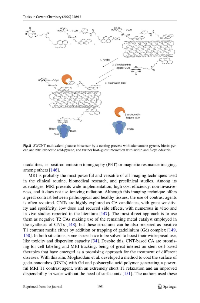
Fig. 8 SWCNT multivalent glucose biosensor by a coating process with adamantane-pyrene, biotin-pyrene and nitrilotriacetic acid-pyrene, and further host–guest interaction with avidin and β-cyclodextrin
195
Reprinted from the journal
Topics in Current Chemistry (2020) 378:15
modalities, as positron emission tomography (PET) or magnetic resonance imaging,
among others [146].
MRI is probably the most powerful and versatile of all imaging techniques used
in the clinical routine, biomedical research, and preclinical studies. Among its
advantages, MRI presents wide implementation, high cost efficiency, non-invasiveness, and it does not use ionizing radiation. Although this imaging technique offers
a great contrast between pathological and healthy tissues, the use of contrast agents
is often required. CNTs are highly explored as CA candidates, with great sensitivity and specificity, low dose and reduced side effects, with numerous in vitro and
in vivo studies reported in the literature [147]. The most direct approach is to use
them as negative T2 CAs making use of the remaining metal catalyst employed in
the synthesis of CNTs [148], but these structures can be also prepared as positive
T1 contrast media either by addition or trapping of gadolinium (Gd) complex [149,
150]. In both situations, some issues have to be solved to boost their widespread use,
like toxicity and dispersion capacity [34]. Despite this, CNT-based CA are promising for cell labeling and MRI tracking, being of great interest on stem cell-based
therapies that have emerged as a promising approach for the treatment of different
diseases. With this aim, Moghaddam et al. developed a method to coat the surface of
gado-nanotubes (GNTs) with Gd and polyacrylic acid polymer generating a powerful MRI T1 contrast agent, with an extremely short T1 relaxation and an improved
dispersibility in water without the need of surfactants [151]. The authors used these
Fig. 8 SWCNT multivalent glucose biosensor by a coating process with adamantane-pyrene, biotin-pyrene and nitrilotriacetic acid-pyrene, and further host–guest interaction with avidin and β-cyclodextrin
195
Reprinted from the journal