

Topics in Current Chemistry (2020) 378:35
1 3
as cell labeling, enzyme activity measurements, tumor diagnosis and therapy and
anatomical localization and real‐time assessment during surgery [158]. Nanostructures based on fluorescent QDs can be synthesized to provide magnetic properties
to the nanomaterial, thereby creating opportunities for multi-modality biomedical
imaging. Fluorescent QDs exhibiting magnetic susceptibility can be synthesized following four different methodologies: metal doping, covalent conjugation, isocrystal
growth and co-encapsulation or electrostatic assembly. In particular, a large number
of fluorescent imaging/MRI dual-modality imaging nanoprobes combine Gd
3+
or
Mn
2+
ions with QDs. As an example, a dual contrast nanoreagent was developed by
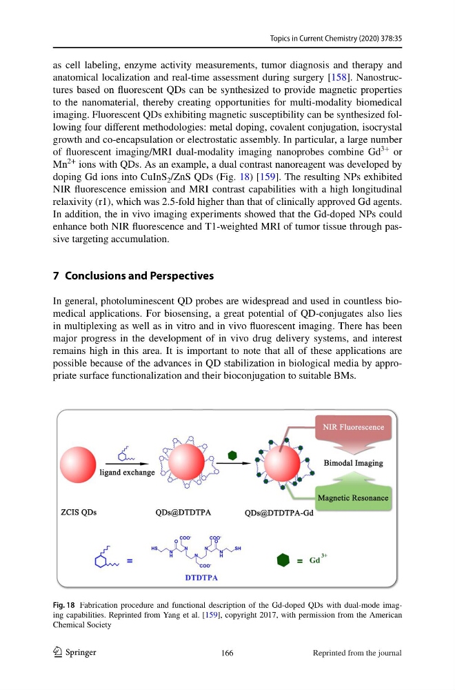
doping Gd ions into CuInS 2 /ZnS QDs (Fig. 18) [159]. The resulting NPs exhibited
NIR fluorescence emission and MRI contrast capabilities with a high longitudinal
relaxivity (r1), which was 2.5-fold higher than that of clinically approved Gd agents.
In addition, the in vivo imaging experiments showed that the Gd-doped NPs could
enhance both NIR fluorescence and T1-weighted MRI of tumor tissue through passive targeting accumulation.
7 Conclusions and Perspectives
In general, photoluminescent QD probes are widespread and used in countless biomedical applications. For biosensing, a great potential of QD-conjugates also lies
in multiplexing as well as in vitro and in vivo fluorescent imaging. There has been
major progress in the development of in vivo drug delivery systems, and interest
remains high in this area. It is important to note that all of these applications are
possible because of the advances in QD stabilization in biological media by appropriate surface functionalization and their bioconjugation to suitable BMs.
Fig. 18 Fabrication procedure and functional description of the Gd-doped QDs with dual-mode imaging capabilities. Reprinted from Yang et al. [159], copyright 2017, with permission from the American
Chemical Society
166
Reprinted from the journal
1 3
as cell labeling, enzyme activity measurements, tumor diagnosis and therapy and
anatomical localization and real‐time assessment during surgery [158]. Nanostructures based on fluorescent QDs can be synthesized to provide magnetic properties
to the nanomaterial, thereby creating opportunities for multi-modality biomedical
imaging. Fluorescent QDs exhibiting magnetic susceptibility can be synthesized following four different methodologies: metal doping, covalent conjugation, isocrystal
growth and co-encapsulation or electrostatic assembly. In particular, a large number
of fluorescent imaging/MRI dual-modality imaging nanoprobes combine Gd
3+
or
Mn
2+
ions with QDs. As an example, a dual contrast nanoreagent was developed by
doping Gd ions into CuInS 2 /ZnS QDs (Fig. 18) [159]. The resulting NPs exhibited
NIR fluorescence emission and MRI contrast capabilities with a high longitudinal
relaxivity (r1), which was 2.5-fold higher than that of clinically approved Gd agents.
In addition, the in vivo imaging experiments showed that the Gd-doped NPs could
enhance both NIR fluorescence and T1-weighted MRI of tumor tissue through passive targeting accumulation.
7 Conclusions and Perspectives
In general, photoluminescent QD probes are widespread and used in countless biomedical applications. For biosensing, a great potential of QD-conjugates also lies
in multiplexing as well as in vitro and in vivo fluorescent imaging. There has been
major progress in the development of in vivo drug delivery systems, and interest
remains high in this area. It is important to note that all of these applications are
possible because of the advances in QD stabilization in biological media by appropriate surface functionalization and their bioconjugation to suitable BMs.
Fig. 18 Fabrication procedure and functional description of the Gd-doped QDs with dual-mode imaging capabilities. Reprinted from Yang et al. [159], copyright 2017, with permission from the American
Chemical Society
166
Reprinted from the journal