

14.2 More Realistic Neuron Models
585
14.2.2 The Hodgkin–Huxley Model
Through their extensive and detailed studies of the electrical properties of squid
neuron membranes (squid axons are particularly big), Alan Hodgkin and Andrew
Huxley described 7 (1952) an electric circuit model of a real neuron’s electrical
activity. Unlike the discrete time sequences used to describe the input and output
of a McCullouch-Pitts artificial neuron, the dynamics of the Hodgkin–Huxley
model neuron are taken continuous in time. Because they used insightful and
consistent mathematical formulations of how ion channels might operate, and used
measurement to pin down what description and parameters to use in the formulation,
their model was impressively successful in matching how a real neuron behaves
when stimulated, earning them a Nobel prize.
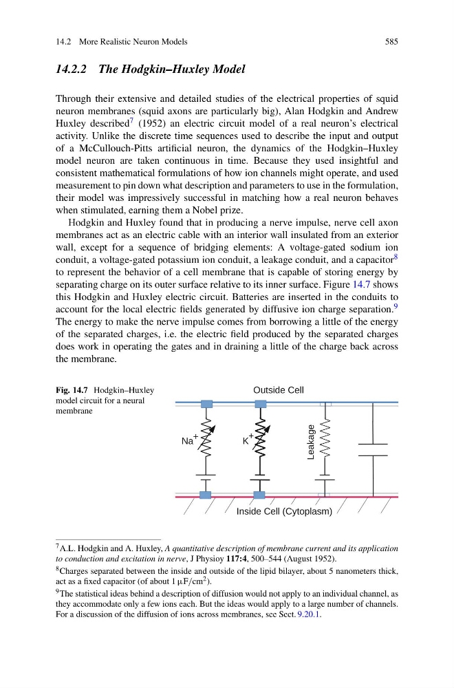
Hodgkin and Huxley found that in producing a nerve impulse, nerve cell axon
membranes act as an electric cable with an interior wall insulated from an exterior
wall, except for a sequence of bridging elements: A voltage-gated sodium ion
conduit, a voltage-gated potassium ion conduit, a leakage conduit, and a capacitor 8
to represent the behavior of a cell membrane that is capable of storing energy by
separating charge on its outer surface relative to its inner surface. Figure 14.7 shows
this Hodgkin and Huxley electric circuit. Batteries are inserted in the conduits to
account for the local electric fields generated by diffusive ion charge separation. 9
The energy to make the nerve impulse comes from borrowing a little of the energy
of the separated charges, i.e. the electric field produced by the separated charges
does work in operating the gates and in draining a little of the charge back across
the membrane.
Fig. 14.7 Hodgkin–Huxley
model circuit for a neural
membrane
7 A.L. Hodgkin and A. Huxley, A quantitative description of membrane current and its application
to conduction and excitation in nerve, J Physioy 117:4, 500–544 (August 1952).
8 Charges separated between the inside and outside of the lipid bilayer, about 5 nanometers thick,
act as a fixed capacitor (of about 1 μF/cm 2 ).
9 The statistical ideas behind a description of diffusion would not apply to an individual channel, as
they accommodate only a few ions each. But the ideas would apply to a large number of channels.
For a discussion of the diffusion of ions across membranes, see Sect. 9.20.1.