

5.22 Medical Applications of Ultrasonics
171
Time (40ms/tick mark)
Doppler shift per unit frequency
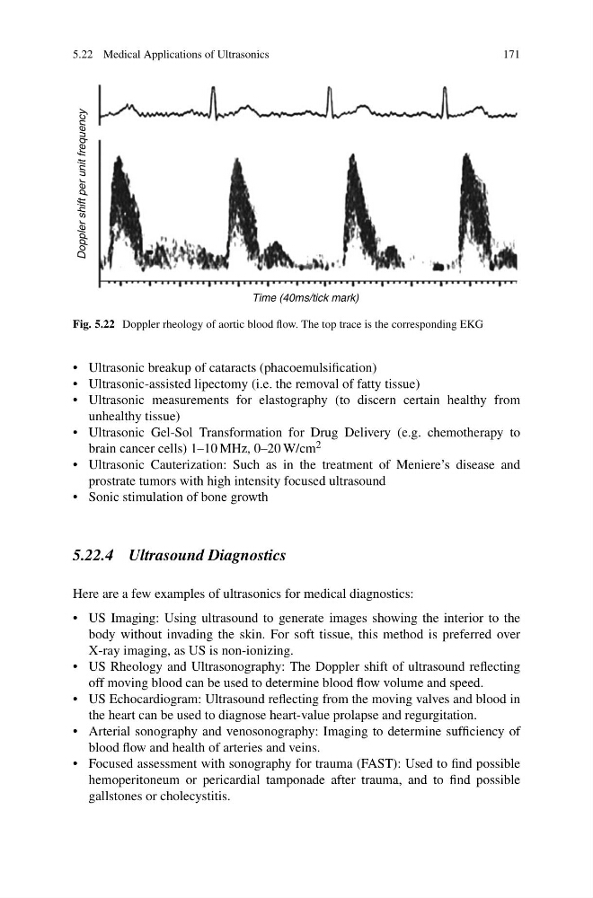
Fig. 5.22 Doppler rheology of aortic blood flow. The top trace is the corresponding EKG
• Ultrasonic breakup of cataracts (phacoemulsification)
• Ultrasonic-assisted lipectomy (i.e. the removal of fatty tissue)
• Ultrasonic measurements for elastography (to discern certain healthy from
unhealthy tissue)
• Ultrasonic Gel-Sol Transformation for Drug Delivery (e.g. chemotherapy to
brain cancer cells) 1–10 MHz, 0–20 W/cm 2
• Ultrasonic Cauterization: Such as in the treatment of Meniere’s disease and
prostrate tumors with high intensity focused ultrasound
• Sonic stimulation of bone growth
5.22.4 Ultrasound Diagnostics
Here are a few examples of ultrasonics for medical diagnostics:
• US Imaging: Using ultrasound to generate images showing the interior to the
body without invading the skin. For soft tissue, this method is preferred over
X-ray imaging, as US is non-ionizing.
• US Rheology and Ultrasonography: The Doppler shift of ultrasound reflecting
off moving blood can be used to determine blood flow volume and speed.
• US Echocardiogram: Ultrasound reflecting from the moving valves and blood in
the heart can be used to diagnose heart-value prolapse and regurgitation.
• Arterial sonography and venosonography: Imaging to determine sufficiency of
blood flow and health of arteries and veins.
• Focused assessment with sonography for trauma (FAST): Used to find possible
hemoperitoneum or pericardial tamponade after trauma, and to find possible
gallstones or cholecystitis.
171
Time (40ms/tick mark)
Doppler shift per unit frequency
Fig. 5.22 Doppler rheology of aortic blood flow. The top trace is the corresponding EKG
• Ultrasonic breakup of cataracts (phacoemulsification)
• Ultrasonic-assisted lipectomy (i.e. the removal of fatty tissue)
• Ultrasonic measurements for elastography (to discern certain healthy from
unhealthy tissue)
• Ultrasonic Gel-Sol Transformation for Drug Delivery (e.g. chemotherapy to
brain cancer cells) 1–10 MHz, 0–20 W/cm 2
• Ultrasonic Cauterization: Such as in the treatment of Meniere’s disease and
prostrate tumors with high intensity focused ultrasound
• Sonic stimulation of bone growth
5.22.4 Ultrasound Diagnostics
Here are a few examples of ultrasonics for medical diagnostics:
• US Imaging: Using ultrasound to generate images showing the interior to the
body without invading the skin. For soft tissue, this method is preferred over
X-ray imaging, as US is non-ionizing.
• US Rheology and Ultrasonography: The Doppler shift of ultrasound reflecting
off moving blood can be used to determine blood flow volume and speed.
• US Echocardiogram: Ultrasound reflecting from the moving valves and blood in
the heart can be used to diagnose heart-value prolapse and regurgitation.
• Arterial sonography and venosonography: Imaging to determine sufficiency of
blood flow and health of arteries and veins.
• Focused assessment with sonography for trauma (FAST): Used to find possible
hemoperitoneum or pericardial tamponade after trauma, and to find possible
gallstones or cholecystitis.