

which is the actual synchrotron component, that accelerates them to the full energy
of the storage ring. At this point the electrons are travelling at around 99.9999% the
speed of light, with an effective mass similar to that of a proton, and they can then be
passed into the storage ring. Unlike a laboratory source, the X-rays are not generated
by colliding with a target, but by making the electrons deviate from their accelerated
trajectory, and as they do so, they have to give off a photon to conserve angular
momentum. The energy of the photon depends on the energy of the electron and the
radius of the turn. The lost energy is replenished using a radio-frequency (RF) cavity.
Due to the combination of bends and straights, the storage ring’s magnetic lattice
forms more of a polygonal shape than a circle.
Synchrotron light sources have undergone an evolution, from particle physics
playthings to large-scale user facilities over the past 70 years. Initially only used for
accelerating particles for collision experiments, the observation of visible light
emanating from a glass vacuum vessel on the 70 meV synchrotron at General
Electric’s Schenectady, NY, labs [27] changed the trajectory of large-scale user
facilities entirely. Within years, scientists came to these particle accelerating sites
and harvested the photons produced by particle physics storage ring operations – this
is considered the first generation of synchrotron light sources, aka, parasitic operation. Beamlines, which direct and condition the light, were built in a generic
fashion – the instrumentation attached was changeable and ad hoc. The second
generation of light sources are those which were designed for the production of
light for scientists, and they were built specifically to produce light in certain regions
of the electromagnetic spectrum [28]. The macromolecular crystallography community were early adopters of synchrotron radiation as the highly collimated beams and
high photon fluxes offered by second-generation machines provided significant
advantages over laboratory-based instrumentation. The beamlines on these early
synchrotrons were often taking light from bending magnets, which produced a
smoothly varying energy spectrum. Bending magnets are characterised by their
critical energy, which is where the total emitted light from the bending magnet can
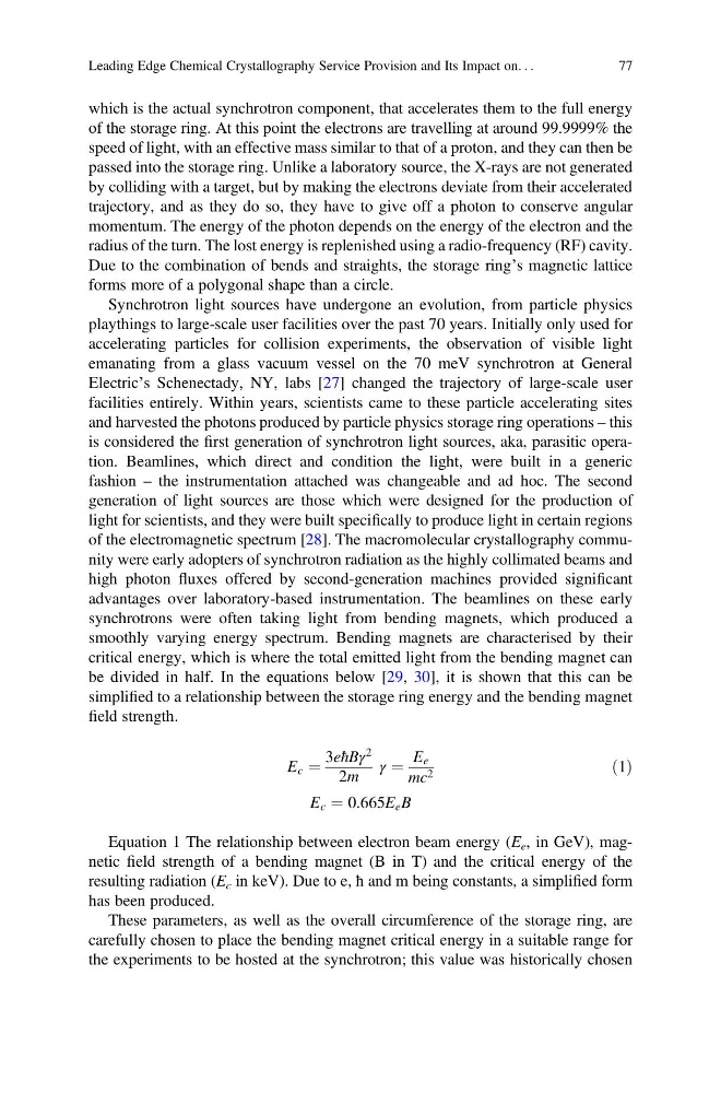
be divided in half. In the equations below [29, 30], it is shown that this can be
simplified to a relationship between the storage ring energy and the bending magnet
field strength.
E c ¼
3eħBγ
2
2m
γ ¼
E e
mc 2
ð1Þ
E c ¼ 0:665E e B
Equation 1 The relationship between electron beam energy (E e , in GeV), magnetic field strength of a bending magnet (B in T) and the critical energy of the
resulting radiation (E c in keV). Due to e, ħ and m being constants, a simplified form
has been produced.
These parameters, as well as the overall circumference of the storage ring, are
carefully chosen to place the bending magnet critical energy in a suitable range for
the experiments to be hosted at the synchrotron; this value was historically chosen
Leading Edge Chemical Crystallography Service Provision and Its Impact on. . .
77
of the storage ring. At this point the electrons are travelling at around 99.9999% the
speed of light, with an effective mass similar to that of a proton, and they can then be
passed into the storage ring. Unlike a laboratory source, the X-rays are not generated
by colliding with a target, but by making the electrons deviate from their accelerated
trajectory, and as they do so, they have to give off a photon to conserve angular
momentum. The energy of the photon depends on the energy of the electron and the
radius of the turn. The lost energy is replenished using a radio-frequency (RF) cavity.
Due to the combination of bends and straights, the storage ring’s magnetic lattice
forms more of a polygonal shape than a circle.
Synchrotron light sources have undergone an evolution, from particle physics
playthings to large-scale user facilities over the past 70 years. Initially only used for
accelerating particles for collision experiments, the observation of visible light
emanating from a glass vacuum vessel on the 70 meV synchrotron at General
Electric’s Schenectady, NY, labs [27] changed the trajectory of large-scale user
facilities entirely. Within years, scientists came to these particle accelerating sites
and harvested the photons produced by particle physics storage ring operations – this
is considered the first generation of synchrotron light sources, aka, parasitic operation. Beamlines, which direct and condition the light, were built in a generic
fashion – the instrumentation attached was changeable and ad hoc. The second
generation of light sources are those which were designed for the production of
light for scientists, and they were built specifically to produce light in certain regions
of the electromagnetic spectrum [28]. The macromolecular crystallography community were early adopters of synchrotron radiation as the highly collimated beams and
high photon fluxes offered by second-generation machines provided significant
advantages over laboratory-based instrumentation. The beamlines on these early
synchrotrons were often taking light from bending magnets, which produced a
smoothly varying energy spectrum. Bending magnets are characterised by their
critical energy, which is where the total emitted light from the bending magnet can
be divided in half. In the equations below [29, 30], it is shown that this can be
simplified to a relationship between the storage ring energy and the bending magnet
field strength.
E c ¼
3eħBγ
2
2m
γ ¼
E e
mc 2
ð1Þ
E c ¼ 0:665E e B
Equation 1 The relationship between electron beam energy (E e , in GeV), magnetic field strength of a bending magnet (B in T) and the critical energy of the
resulting radiation (E c in keV). Due to e, ħ and m being constants, a simplified form
has been produced.
These parameters, as well as the overall circumference of the storage ring, are
carefully chosen to place the bending magnet critical energy in a suitable range for
the experiments to be hosted at the synchrotron; this value was historically chosen
Leading Edge Chemical Crystallography Service Provision and Its Impact on. . .
77