

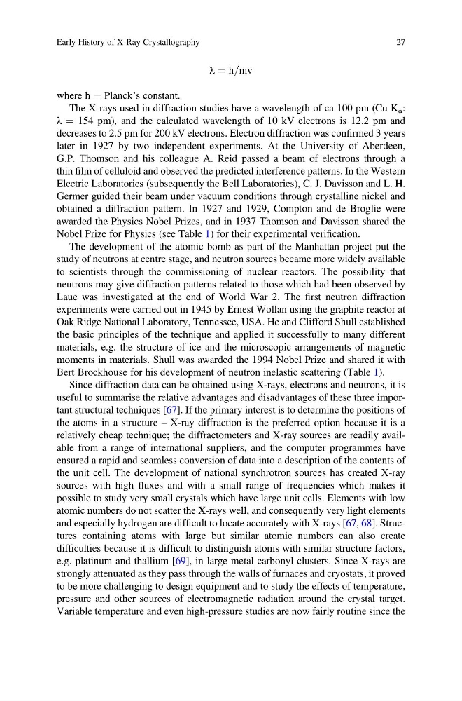
λ ¼ h=mv
where h ¼ Planck’s constant.
The X-rays used in diffraction studies have a wavelength of ca 100 pm (Cu K α :
λ ¼ 154 pm), and the calculated wavelength of 10 kV electrons is 12.2 pm and
decreases to 2.5 pm for 200 kV electrons. Electron diffraction was confirmed 3 years
later in 1927 by two independent experiments. At the University of Aberdeen,
G.P. Thomson and his colleague A. Reid passed a beam of electrons through a
thin film of celluloid and observed the predicted interference patterns. In the Western
Electric Laboratories (subsequently the Bell Laboratories), C. J. Davisson and L. H.
Germer guided their beam under vacuum conditions through crystalline nickel and
obtained a diffraction pattern. In 1927 and 1929, Compton and de Broglie were
awarded the Physics Nobel Prizes, and in 1937 Thomson and Davisson shared the
Nobel Prize for Physics (see Table 1) for their experimental verification.
The development of the atomic bomb as part of the Manhattan project put the
study of neutrons at centre stage, and neutron sources became more widely available
to scientists through the commissioning of nuclear reactors. The possibility that
neutrons may give diffraction patterns related to those which had been observed by
Laue was investigated at the end of World War 2. The first neutron diffraction
experiments were carried out in 1945 by Ernest Wollan using the graphite reactor at
Oak Ridge National Laboratory, Tennessee, USA. He and Clifford Shull established
the basic principles of the technique and applied it successfully to many different
materials, e.g. the structure of ice and the microscopic arrangements of magnetic
moments in materials. Shull was awarded the 1994 Nobel Prize and shared it with
Bert Brockhouse for his development of neutron inelastic scattering (Table 1).
Since diffraction data can be obtained using X-rays, electrons and neutrons, it is
useful to summarise the relative advantages and disadvantages of these three important structural techniques [67]. If the primary interest is to determine the positions of
the atoms in a structure – X-ray diffraction is the preferred option because it is a
relatively cheap technique; the diffractometers and X-ray sources are readily available from a range of international suppliers, and the computer programmes have
ensured a rapid and seamless conversion of data into a description of the contents of
the unit cell. The development of national synchrotron sources has created X-ray
sources with high fluxes and with a small range of frequencies which makes it
possible to study very small crystals which have large unit cells. Elements with low
atomic numbers do not scatter the X-rays well, and consequently very light elements
and especially hydrogen are difficult to locate accurately with X-rays [67, 68]. Structures containing atoms with large but similar atomic numbers can also create
difficulties because it is difficult to distinguish atoms with similar structure factors,
e.g. platinum and thallium [69], in large metal carbonyl clusters. Since X-rays are
strongly attenuated as they pass through the walls of furnaces and cryostats, it proved
to be more challenging to design equipment and to study the effects of temperature,
pressure and other sources of electromagnetic radiation around the crystal target.
Variable temperature and even high-pressure studies are now fairly routine since the
Early History of X-Ray Crystallography
27
where h ¼ Planck’s constant.
The X-rays used in diffraction studies have a wavelength of ca 100 pm (Cu K α :
λ ¼ 154 pm), and the calculated wavelength of 10 kV electrons is 12.2 pm and
decreases to 2.5 pm for 200 kV electrons. Electron diffraction was confirmed 3 years
later in 1927 by two independent experiments. At the University of Aberdeen,
G.P. Thomson and his colleague A. Reid passed a beam of electrons through a
thin film of celluloid and observed the predicted interference patterns. In the Western
Electric Laboratories (subsequently the Bell Laboratories), C. J. Davisson and L. H.
Germer guided their beam under vacuum conditions through crystalline nickel and
obtained a diffraction pattern. In 1927 and 1929, Compton and de Broglie were
awarded the Physics Nobel Prizes, and in 1937 Thomson and Davisson shared the
Nobel Prize for Physics (see Table 1) for their experimental verification.
The development of the atomic bomb as part of the Manhattan project put the
study of neutrons at centre stage, and neutron sources became more widely available
to scientists through the commissioning of nuclear reactors. The possibility that
neutrons may give diffraction patterns related to those which had been observed by
Laue was investigated at the end of World War 2. The first neutron diffraction
experiments were carried out in 1945 by Ernest Wollan using the graphite reactor at
Oak Ridge National Laboratory, Tennessee, USA. He and Clifford Shull established
the basic principles of the technique and applied it successfully to many different
materials, e.g. the structure of ice and the microscopic arrangements of magnetic
moments in materials. Shull was awarded the 1994 Nobel Prize and shared it with
Bert Brockhouse for his development of neutron inelastic scattering (Table 1).
Since diffraction data can be obtained using X-rays, electrons and neutrons, it is
useful to summarise the relative advantages and disadvantages of these three important structural techniques [67]. If the primary interest is to determine the positions of
the atoms in a structure – X-ray diffraction is the preferred option because it is a
relatively cheap technique; the diffractometers and X-ray sources are readily available from a range of international suppliers, and the computer programmes have
ensured a rapid and seamless conversion of data into a description of the contents of
the unit cell. The development of national synchrotron sources has created X-ray
sources with high fluxes and with a small range of frequencies which makes it
possible to study very small crystals which have large unit cells. Elements with low
atomic numbers do not scatter the X-rays well, and consequently very light elements
and especially hydrogen are difficult to locate accurately with X-rays [67, 68]. Structures containing atoms with large but similar atomic numbers can also create
difficulties because it is difficult to distinguish atoms with similar structure factors,
e.g. platinum and thallium [69], in large metal carbonyl clusters. Since X-rays are
strongly attenuated as they pass through the walls of furnaces and cryostats, it proved
to be more challenging to design equipment and to study the effects of temperature,
pressure and other sources of electromagnetic radiation around the crystal target.
Variable temperature and even high-pressure studies are now fairly routine since the
Early History of X-Ray Crystallography
27