

2.4 Sub-picosecond and XFEL Methodologies
The interest in studying chemical and biological processes with shorter and shorter
lifetimes using time-resolved crystallographic techniques continues to grow as
technological advances make the picosecond and sub-picosecond time regimes
accessible. The structure of the synchrotron beam itself can help with these developments. The relativistic effects on the electrons circulating the storage ring at
speeds approaching that of light mean that they orbit the ring in discrete bunches
[22]. Thus, the synchrotron radiation produced is naturally pulsed with a repetition
rate that is determined by the period of the electron orbit around the ring. For thirdgeneration synchrotrons, this is typically in the nanosecond to picosecond time
regime. Therefore, pump-probe time-resolved experiments on species with lifetimes
within this timeframe can be carried out without the need for a mechanical or
electronic shutter to pulse the X-rays, and the laser repetition rate is synchronised
with the repetition rate of the storage ring [40].
In order to cover all dynamic chemical processes, the ultimate aim must be to use
photocrystallographic methods to investigate the initial stages of a chemical reaction
that occur on femtosecond timescales [41]. The study of species with
sub-picosecond lifetimes requires the development of “single-shot” diffraction
methods where the whole diffraction pattern of the crystal is obtained in one X-ray
pulse. Here the flux of the X-ray pulse needs to be extremely high in order to achieve
a measurable pattern. Laue techniques provide sufficient flux to obtain the result, but
the development of X-ray free-electron lasers (XFELs) with several orders of
magnitude more flux than the most powerful synchrotrons is the obvious tool to
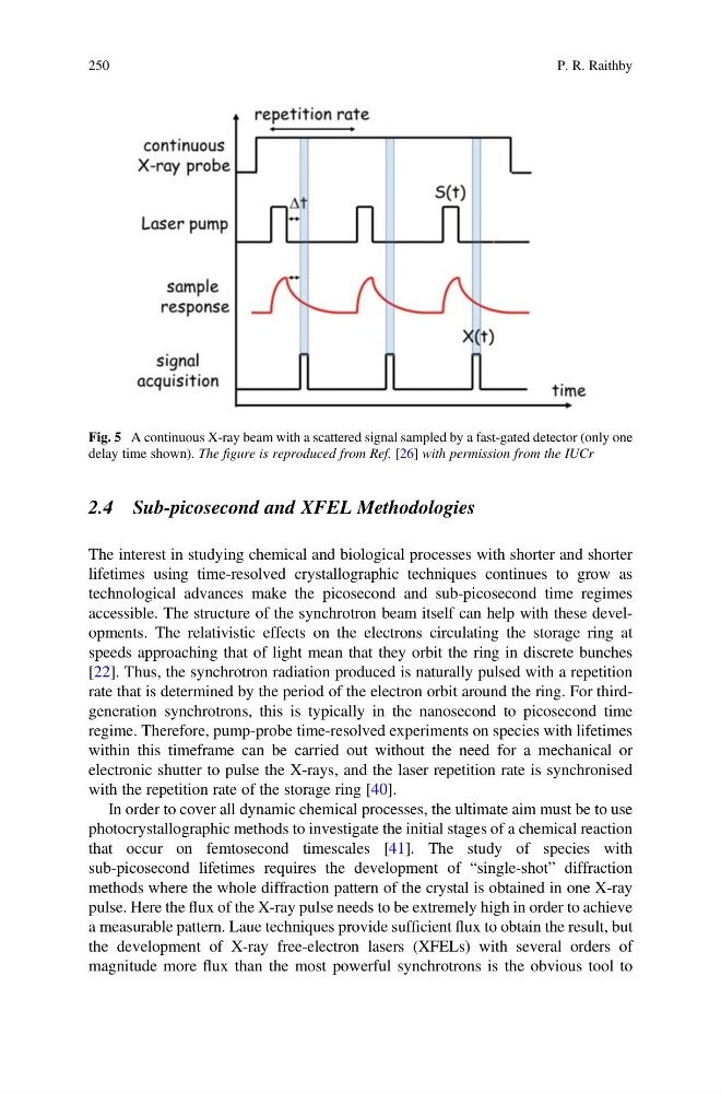
Fig. 5 A continuous X-ray beam with a scattered signal sampled by a fast-gated detector (only one
delay time shown). The figure is reproduced from Ref. [26] with permission from the IUCr
250
P. R. Raithby
The interest in studying chemical and biological processes with shorter and shorter
lifetimes using time-resolved crystallographic techniques continues to grow as
technological advances make the picosecond and sub-picosecond time regimes
accessible. The structure of the synchrotron beam itself can help with these developments. The relativistic effects on the electrons circulating the storage ring at
speeds approaching that of light mean that they orbit the ring in discrete bunches
[22]. Thus, the synchrotron radiation produced is naturally pulsed with a repetition
rate that is determined by the period of the electron orbit around the ring. For thirdgeneration synchrotrons, this is typically in the nanosecond to picosecond time
regime. Therefore, pump-probe time-resolved experiments on species with lifetimes
within this timeframe can be carried out without the need for a mechanical or
electronic shutter to pulse the X-rays, and the laser repetition rate is synchronised
with the repetition rate of the storage ring [40].
In order to cover all dynamic chemical processes, the ultimate aim must be to use
photocrystallographic methods to investigate the initial stages of a chemical reaction
that occur on femtosecond timescales [41]. The study of species with
sub-picosecond lifetimes requires the development of “single-shot” diffraction
methods where the whole diffraction pattern of the crystal is obtained in one X-ray
pulse. Here the flux of the X-ray pulse needs to be extremely high in order to achieve
a measurable pattern. Laue techniques provide sufficient flux to obtain the result, but
the development of X-ray free-electron lasers (XFELs) with several orders of
magnitude more flux than the most powerful synchrotrons is the obvious tool to
Fig. 5 A continuous X-ray beam with a scattered signal sampled by a fast-gated detector (only one
delay time shown). The figure is reproduced from Ref. [26] with permission from the IUCr
250
P. R. Raithby