

2.3 Laue Methods
The time-resolved experiments described so far have used monochromatic X-ray
radiation. An alternative way of overcoming the limited flux in short-duration timeresolved crystallographic experiments is to change from monochromatic radiation to
the Laue method where polychromatic X-ray radiation is used [29, 30]. The main
advantage of the Laue method is the broader energy range used, compared with the
narrow beam from a monochromator. This results in a substantial increase in
intensity. The method used in both molecular [31] and macromolecular [32] crystallographic experiments is often called the pink-Laue method as “pink” indicates
quite a small range of wavelengths as opposed to all the wavelengths available in the
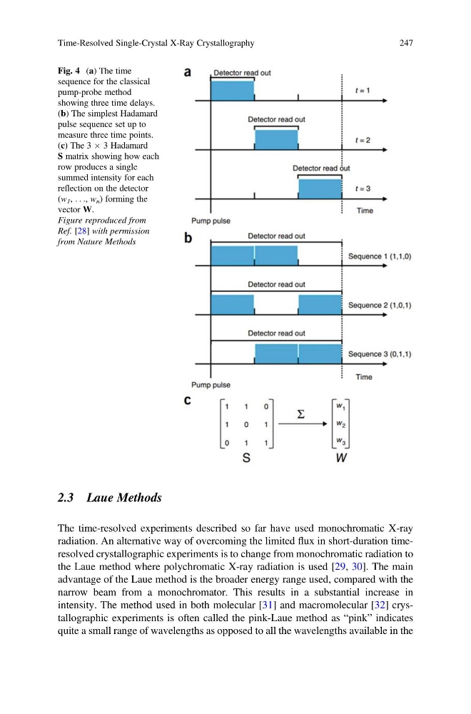
Fig. 4 (a) The time
sequence for the classical
pump-probe method
showing three time delays.
(b) The simplest Hadamard
pulse sequence set up to
measure three time points.
(c) The 3 Â 3 Hadamard
S matrix showing how each
row produces a single
summed intensity for each
reflection on the detector
(w 1 , . . ., w n ) forming the
vector W.
Figure reproduced from
Ref. [28] with permission
from Nature Methods
Time-Resolved Single-Crystal X-Ray Crystallography
247
The time-resolved experiments described so far have used monochromatic X-ray
radiation. An alternative way of overcoming the limited flux in short-duration timeresolved crystallographic experiments is to change from monochromatic radiation to
the Laue method where polychromatic X-ray radiation is used [29, 30]. The main
advantage of the Laue method is the broader energy range used, compared with the
narrow beam from a monochromator. This results in a substantial increase in
intensity. The method used in both molecular [31] and macromolecular [32] crystallographic experiments is often called the pink-Laue method as “pink” indicates
quite a small range of wavelengths as opposed to all the wavelengths available in the
Fig. 4 (a) The time
sequence for the classical
pump-probe method
showing three time delays.
(b) The simplest Hadamard
pulse sequence set up to
measure three time points.
(c) The 3 Â 3 Hadamard
S matrix showing how each
row produces a single
summed intensity for each
reflection on the detector
(w 1 , . . ., w n ) forming the
vector W.
Figure reproduced from
Ref. [28] with permission
from Nature Methods
Time-Resolved Single-Crystal X-Ray Crystallography
247