

science to be conducted routinely in a laboratory environment. Its simple design and
ease of use has resulted in it becoming the workhorse of many high-pressure
laboratories. The original design of the DAC used beryllium discs as backing plates
for the diamonds to sit on. The beryllium plates provided a mechanically strong
material whilst being relatively transparent to X-rays due to the low electron count of
beryllium. The backing discs produced diffraction rings themselves which increased
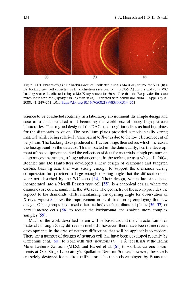
the background on the detector. This impacted on the data quality, but the development of the equipment enabled the collection of data for materials at high pressure on
a laboratory instrument, a huge advancement in the technique as a whole. In 2004,
Boehler and De Hantsetters developed a new design of diamonds and tungsten
carbide backing seat that was strong enough to support the diamonds during
compression but provided a large enough opening angle that the diffraction data
were not absorbed by the WC seats [54]. Their design, which has since been
incorporated into a Merrill-Bassett-type cell [55], is a canonical design where the
diamonds are countersunk into the WC seat. The geometry of the set-up provides the
support to the diamonds whilst maintaining the opening angle for observation of
X-rays. Figure 5 shows the improvement in the diffraction by employing this new
design. Other groups have used other methods such as diamond plates [56, 57] or
beryllium-free cells [58] to reduce the background and analyse more complex
samples [59].
Much of the work described herein will be based around the characterisation of
materials through X-ray diffraction methods; however, there have been some recent
developments in the area of neutron diffraction that will be applicable to readers.
There are a number of designs of neutron cell that have been developed recently by
Grzechnik et al. [60], to work with ‘hot’ neutrons (λ ¼ 1 Å) at HEiDi at the Heinz
Maier-Leibnitz Zentrum (MLZ), and Haberl et al. [61] to work at various instruments at Oak Ridge Laboratory’s Spallation Neutron Source; however, these cells
are solely designed for neutron diffraction. The methods employed by Binns and
Fig. 5 CCD images of (a) a Be backing-seat cell collected using a Mo X-ray source for 60 s, (b) a
Be backing-seat cell collected with synchrotron radiation (λ ¼ 0.6755 Å) for 1 s and (c) a WC
backing-seat cell collected using a Mo X-ray source for 60 s. Note that the Be powder lines are
much more textured (‘spotty’) in (b) than in (a). Reprinted with permission from J. Appl. Cryst.,
2008, 41, 249–251, DOI: https://doi.org/10.1107/S0021889808000514 [55]
154
S. A. Moggach and I. D. H. Oswald
ease of use has resulted in it becoming the workhorse of many high-pressure
laboratories. The original design of the DAC used beryllium discs as backing plates
for the diamonds to sit on. The beryllium plates provided a mechanically strong
material whilst being relatively transparent to X-rays due to the low electron count of
beryllium. The backing discs produced diffraction rings themselves which increased
the background on the detector. This impacted on the data quality, but the development of the equipment enabled the collection of data for materials at high pressure on
a laboratory instrument, a huge advancement in the technique as a whole. In 2004,
Boehler and De Hantsetters developed a new design of diamonds and tungsten
carbide backing seat that was strong enough to support the diamonds during
compression but provided a large enough opening angle that the diffraction data
were not absorbed by the WC seats [54]. Their design, which has since been
incorporated into a Merrill-Bassett-type cell [55], is a canonical design where the
diamonds are countersunk into the WC seat. The geometry of the set-up provides the
support to the diamonds whilst maintaining the opening angle for observation of
X-rays. Figure 5 shows the improvement in the diffraction by employing this new
design. Other groups have used other methods such as diamond plates [56, 57] or
beryllium-free cells [58] to reduce the background and analyse more complex
samples [59].
Much of the work described herein will be based around the characterisation of
materials through X-ray diffraction methods; however, there have been some recent
developments in the area of neutron diffraction that will be applicable to readers.
There are a number of designs of neutron cell that have been developed recently by
Grzechnik et al. [60], to work with ‘hot’ neutrons (λ ¼ 1 Å) at HEiDi at the Heinz
Maier-Leibnitz Zentrum (MLZ), and Haberl et al. [61] to work at various instruments at Oak Ridge Laboratory’s Spallation Neutron Source; however, these cells
are solely designed for neutron diffraction. The methods employed by Binns and
Fig. 5 CCD images of (a) a Be backing-seat cell collected using a Mo X-ray source for 60 s, (b) a
Be backing-seat cell collected with synchrotron radiation (λ ¼ 0.6755 Å) for 1 s and (c) a WC
backing-seat cell collected using a Mo X-ray source for 60 s. Note that the Be powder lines are
much more textured (‘spotty’) in (b) than in (a). Reprinted with permission from J. Appl. Cryst.,
2008, 41, 249–251, DOI: https://doi.org/10.1107/S0021889808000514 [55]
154
S. A. Moggach and I. D. H. Oswald