

42
3 Experimental Section
589.44 nm (sodium D line) in a cell with 100 mm path length. [α]
20
D values
were reported in 10
−1 deg cm
2 g
−1 . X-Ray Analysis: The X-ray intensity
data were measured on a Bruker D8 Venture diffractometer equipped with
multilayer monochromator, Cu-K α INCOATEC micro focus sealed tube and
Oxford cooling device. The structure was solved by direct methods and
refined by full-matrix least-squares techniques. Non-hydrogen atoms were
refined with anisotropic displacement parameters. Hydrogen atoms were inserted at calculated positions and refined with a riding model. The following
software was used: Bruker SAINT software package using a narrow-frame
algorithm for frame integration,
[55] SADABS for absorption correction,
[56]
OLEX2 for structure solution, refinement, molecular diagrams and graphical
user-interface,
[57] ShelXle for refinement and graphical user-interface,
[58]
SHELXS-2013 for structure solution,
[59] SHELXL-2013 for refinement,
[58]
Platon for symmetry check,
[60] mercury for image compilation.
[61]
3.2 Experimental Procedures
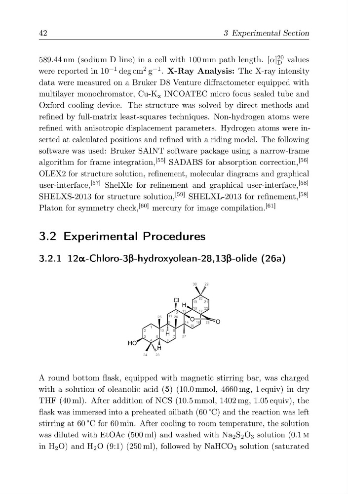
3.2.1 12α-Chloro-3β-hydroxyolean-28,13β-olide (26a)
HO
H
H
H
Cl
O
O
1
2
3
4
5
6
7
8
9
10
11
12 13
14
15
16
17
18
19
20
21
22
28
24
23
25
26
29
27
30
A round bottom flask, equipped with magnetic stirring bar, was charged
with a solution of oleanolic acid (5) (10.0 mmol, 4660 mg, 1 equiv) in dry
THF (40 ml). After addition of NCS (10.5 mmol, 1402 mg, 1.05 equiv), the
flask was immersed into a preheated oilbath (60 °C) and the reaction was left
stirring at 60 °C for 60 min. After cooling to room temperature, the solution
was diluted with EtOAc (500 ml) and washed with Na 2 S 2 O 3 solution (0.1 m
in H 2 O) and H 2 O (9:1) (250 ml), followed by NaHCO 3 solution (saturated
3 Experimental Section
589.44 nm (sodium D line) in a cell with 100 mm path length. [α]
20
D values
were reported in 10
−1 deg cm
2 g
−1 . X-Ray Analysis: The X-ray intensity
data were measured on a Bruker D8 Venture diffractometer equipped with
multilayer monochromator, Cu-K α INCOATEC micro focus sealed tube and
Oxford cooling device. The structure was solved by direct methods and
refined by full-matrix least-squares techniques. Non-hydrogen atoms were
refined with anisotropic displacement parameters. Hydrogen atoms were inserted at calculated positions and refined with a riding model. The following
software was used: Bruker SAINT software package using a narrow-frame
algorithm for frame integration,
[55] SADABS for absorption correction,
[56]
OLEX2 for structure solution, refinement, molecular diagrams and graphical
user-interface,
[57] ShelXle for refinement and graphical user-interface,
[58]
SHELXS-2013 for structure solution,
[59] SHELXL-2013 for refinement,
[58]
Platon for symmetry check,
[60] mercury for image compilation.
[61]
3.2 Experimental Procedures
3.2.1 12α-Chloro-3β-hydroxyolean-28,13β-olide (26a)
HO
H
H
H
Cl
O
O
1
2
3
4
5
6
7
8
9
10
11
12 13
14
15
16
17
18
19
20
21
22
28
24
23
25
26
29
27
30
A round bottom flask, equipped with magnetic stirring bar, was charged
with a solution of oleanolic acid (5) (10.0 mmol, 4660 mg, 1 equiv) in dry
THF (40 ml). After addition of NCS (10.5 mmol, 1402 mg, 1.05 equiv), the
flask was immersed into a preheated oilbath (60 °C) and the reaction was left
stirring at 60 °C for 60 min. After cooling to room temperature, the solution
was diluted with EtOAc (500 ml) and washed with Na 2 S 2 O 3 solution (0.1 m
in H 2 O) and H 2 O (9:1) (250 ml), followed by NaHCO 3 solution (saturated