

11 Application of Accelerators and Storage Rings
725
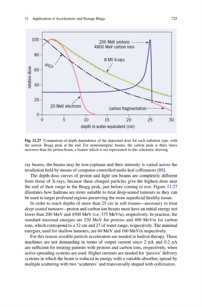
Fig. 11.27 Comparison of depth dependence of the deposited dose for each radiation type, with
the narrow Bragg peak at the end. For monoenergetic beams, the carbon peak is three times
narrower than the proton beam, a feature which is not represented in this schematic drawing
ray beams; the beams may be non-coplanar and their intensity is varied across the
irradiation field by means of computer-controlled multi-leaf collimators [89].
The depth-dose curves of proton and light ion beams are completely different
from those of X-rays, because these charged particles give the highest dose near
the end of their range in the Bragg peak, just before coming to rest. Figure 11.27
illustrates how hadrons are more suitable to treat deep-seated tumours as they can
be used to target profound regions preserving the more superficial healthy tissue.
In order to reach depths of more than 25 cm in soft tissues—necessary to treat
deep-seated tumours—proton and carbon ion beams must have an initial energy not
lower than 200 MeV and 4500 MeV (i.e. 375 MeV/u), respectively. In practice, the
standard maximal energies are 230 MeV for protons and 400 MeV/u for carbon
ions, which correspond to a 32 cm and 27 of water range, respectively. The minimal
energies, used for shallow tumours, are 60 MeV and 100 MeV/u respectively.
For this reason sizeable particle accelerators are needed in hadron therapy. These
machines are not demanding in terms of output current since 2 nA and 0.2 nA
are sufficient for treating patients with protons and carbon ions, respectively, when
active spreading systems are used. Higher currents are needed for ‘passive’ delivery
systems in which the beam is reduced in energy with a variable absorber, spread by
multiple scattering with two ‘scatterers’ and transversally shaped with collimators.