

Fig. 4
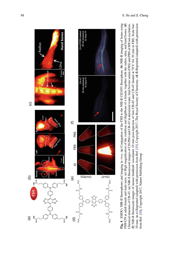
SMDCs NIR-II
fluorophores and imaging in vivo. (a) Conjugation of the FSH to the NIR-II CH1055
fluorophore. (b) NIR-II imaging of bones using
FSH-CH in adult female and male mice. (c) High magnification of the vertebrae and thighbone of a female, together with the radius, ulna, and hand bones. (d)
Chemical structure of CH-4T. (e) NIR-II
fluorescent images of CH-PEG and CH-4T in deionized water, fetal bovine serum (FBS) and PBS at 808 nm excitation.
(f) NIR-II
fluorescent images of mouse hindlimb vasculature 10 min post-injection of free CH-4T and CH-4T heated at 70
C for 10 min in FBS. Scale bar:
10 mm. (a–c) Reprinted (adapted) with permission from Ref. [15], Copyright 2017, The Royal Society of Chemistry. (d–f) Reprinted (adapted) with permission
from Ref. [10], Copyright 2017, Nature Publishing Group
88
S. He and Z. Cheng
SMDCs NIR-II
fluorophores and imaging in vivo. (a) Conjugation of the FSH to the NIR-II CH1055
fluorophore. (b) NIR-II imaging of bones using
FSH-CH in adult female and male mice. (c) High magnification of the vertebrae and thighbone of a female, together with the radius, ulna, and hand bones. (d)
Chemical structure of CH-4T. (e) NIR-II
fluorescent images of CH-PEG and CH-4T in deionized water, fetal bovine serum (FBS) and PBS at 808 nm excitation.
(f) NIR-II
fluorescent images of mouse hindlimb vasculature 10 min post-injection of free CH-4T and CH-4T heated at 70
C for 10 min in FBS. Scale bar:
10 mm. (a–c) Reprinted (adapted) with permission from Ref. [15], Copyright 2017, The Royal Society of Chemistry. (d–f) Reprinted (adapted) with permission
from Ref. [10], Copyright 2017, Nature Publishing Group
88
S. He and Z. Cheng