

Fig. 10
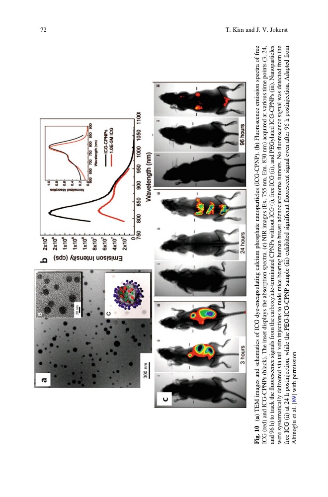
(a) TEM images and schematics of ICG dye-encapsulating calcium phosphate nanoparticles (ICG-CPNP). (b) Fluorescence emission spectra of free
ICG (red) and ICG-CPNPs (black). The inset displays the absorption spectra. (c) NIR images (Ex. 755 nm, Em. 830 nm) acquired at various time points (3, 24,
and 96 h) to track the
fluorescence signals from the carboxylate-terminated CPNPs without ICG (i), free ICG (ii), and PEGylated ICG-CPNPs (iii). Nanoparticles
were systematically delivered via tail vein injections to nude mice bearing human breast adenocarcinoma tumors. No
fluorescence signal was detected from the
free ICG (ii) at 24 h postinjection, while the PEG-ICG-CPNP sample (iii) exhibited significant
fluorescent signal even after 96 h postinjection. Adapted from
Altinoglu et al. [89] with permission
72
T. Kim and J. V. Jokerst
(a) TEM images and schematics of ICG dye-encapsulating calcium phosphate nanoparticles (ICG-CPNP). (b) Fluorescence emission spectra of free
ICG (red) and ICG-CPNPs (black). The inset displays the absorption spectra. (c) NIR images (Ex. 755 nm, Em. 830 nm) acquired at various time points (3, 24,
and 96 h) to track the
fluorescence signals from the carboxylate-terminated CPNPs without ICG (i), free ICG (ii), and PEGylated ICG-CPNPs (iii). Nanoparticles
were systematically delivered via tail vein injections to nude mice bearing human breast adenocarcinoma tumors. No
fluorescence signal was detected from the
free ICG (ii) at 24 h postinjection, while the PEG-ICG-CPNP sample (iii) exhibited significant
fluorescent signal even after 96 h postinjection. Adapted from
Altinoglu et al. [89] with permission
72
T. Kim and J. V. Jokerst