

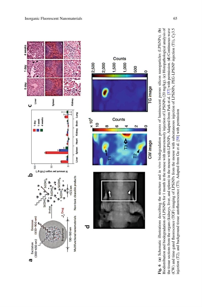
Fig. 6
(a) Schematic illustrations describing the structure and in vivo biodegradation process of luminescent porous silicon nanoparticles (LPSiNPs). (b)
Biodistribution and biodegradation of LPSiNPs for 1 month in the mouse with intravenously injection of LPSiNPs (20 mg/kg). (c) Histopathological analysis of
the tissue sections from the organs (kidneys, liver, and spleen) in the mouse with LPSiNPs. Adapted from Park et al. [57] with permission. (d) Continuous-wave
(CW) and time-gated
fluorescence (TGF) imaging of LPSiNPs from the mouse with subcutaneous injection of LPSiNPs; PEG-LPSiNP injection (T1), Cy3.5
injection (T2), and background tissue autofluorescence (T3). Adapted from Gu et al. [58] with permission
Inorganic Fluorescent Nanomaterials
65
(a) Schematic illustrations describing the structure and in vivo biodegradation process of luminescent porous silicon nanoparticles (LPSiNPs). (b)
Biodistribution and biodegradation of LPSiNPs for 1 month in the mouse with intravenously injection of LPSiNPs (20 mg/kg). (c) Histopathological analysis of
the tissue sections from the organs (kidneys, liver, and spleen) in the mouse with LPSiNPs. Adapted from Park et al. [57] with permission. (d) Continuous-wave
(CW) and time-gated
fluorescence (TGF) imaging of LPSiNPs from the mouse with subcutaneous injection of LPSiNPs; PEG-LPSiNP injection (T1), Cy3.5
injection (T2), and background tissue autofluorescence (T3). Adapted from Gu et al. [58] with permission
Inorganic Fluorescent Nanomaterials
65