

[4]. Remarkably, both of these systems can still utilize the same ~400 nm
excitation.
Consequently, by capitalizing on the striking optical features of QDs (e.g., stale
and sharp emission, spectral nature of emitted photons), Nie and coworkers reported
the first study of QDs for targeting and spectral optical imaging in animal models
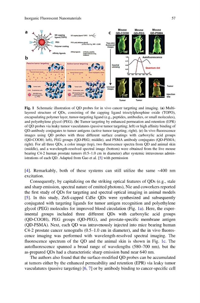
[5]. In this study, ZnS-capped CdSe QDs were synthesized and subsequently
conjugated with targeting ligands for tumor antigen recognition and polyethylene
glycol (PEG) molecules for improved blood circulation (Fig. 1a). Here, the experimental groups included three different QDs with carboxylic acid groups
(QD-COOH), PEG groups (QD-PEG), and prostate-specific membrane antigen
(QD-PSMA). Next, each QD was intravenously injected into mice bearing human
C4-2 prostate cancer xenografts (0.5–1.0 cm in diameter), and the in vivo fluorescence imaging was performed with wavelength-resolved spectral imaging. The
fluorescence spectrum of the QD and the animal skin is shown in Fig. 1c. The
autofluorescence spanned a broad range of wavelengths (580–700 nm), but the
as-prepared QDs had a characteristic sharp emission band near 640 nm.
The authors also found that the surface-modified QD probes can be accumulated
at tumors either by the enhanced permeability and retention (EPR) via leaky tumor
vasculatures (passive targeting) [6, 7] or by antibody binding to cancer-specific cell
Fig. 1 Schematic illustration of QD probes for in vivo cancer targeting and imaging. (a) Multilayered structure of QDs, consisting of the capping ligand trioctylphosphine oxide (TOPO),
encapsulating polymer layer, tumor-targeting ligand (e.g., peptides, antibodies, or small molecules),
and polyethylene glycol (PEG). (b) Tumor targeting by enhanced permeation and retention (EPR)
of QD probes via leaky tumor vasculatures (passive tumor targeting; left) or high affinity binding of
QD-antibody conjugates to tumor antigens (active tumor targeting; right). (c) In vivo fluorescence
images using QD probes with three different surface coatings with carboxylic acid groups
(QD-COOH; left), PEG groups (QD-PEG; middle), and PSMA antibody conjugates (QD-PSMA;
right). For all three QDs, a color image (top), two fluorescence spectra from QD and animal skin
(middle), and a wavelength-resolved spectral image (bottom) were obtained from the live mouse
bearing C4-2 human prostate tumors (0.5–1.0 cm in diameter) after systemic intravenous administrations of each QD. Adapted from Gao et al. [5] with permission
Inorganic Fluorescent Nanomaterials
57
excitation.
Consequently, by capitalizing on the striking optical features of QDs (e.g., stale
and sharp emission, spectral nature of emitted photons), Nie and coworkers reported
the first study of QDs for targeting and spectral optical imaging in animal models
[5]. In this study, ZnS-capped CdSe QDs were synthesized and subsequently
conjugated with targeting ligands for tumor antigen recognition and polyethylene
glycol (PEG) molecules for improved blood circulation (Fig. 1a). Here, the experimental groups included three different QDs with carboxylic acid groups
(QD-COOH), PEG groups (QD-PEG), and prostate-specific membrane antigen
(QD-PSMA). Next, each QD was intravenously injected into mice bearing human
C4-2 prostate cancer xenografts (0.5–1.0 cm in diameter), and the in vivo fluorescence imaging was performed with wavelength-resolved spectral imaging. The
fluorescence spectrum of the QD and the animal skin is shown in Fig. 1c. The
autofluorescence spanned a broad range of wavelengths (580–700 nm), but the
as-prepared QDs had a characteristic sharp emission band near 640 nm.
The authors also found that the surface-modified QD probes can be accumulated
at tumors either by the enhanced permeability and retention (EPR) via leaky tumor
vasculatures (passive targeting) [6, 7] or by antibody binding to cancer-specific cell
Fig. 1 Schematic illustration of QD probes for in vivo cancer targeting and imaging. (a) Multilayered structure of QDs, consisting of the capping ligand trioctylphosphine oxide (TOPO),
encapsulating polymer layer, tumor-targeting ligand (e.g., peptides, antibodies, or small molecules),
and polyethylene glycol (PEG). (b) Tumor targeting by enhanced permeation and retention (EPR)
of QD probes via leaky tumor vasculatures (passive tumor targeting; left) or high affinity binding of
QD-antibody conjugates to tumor antigens (active tumor targeting; right). (c) In vivo fluorescence
images using QD probes with three different surface coatings with carboxylic acid groups
(QD-COOH; left), PEG groups (QD-PEG; middle), and PSMA antibody conjugates (QD-PSMA;
right). For all three QDs, a color image (top), two fluorescence spectra from QD and animal skin
(middle), and a wavelength-resolved spectral image (bottom) were obtained from the live mouse
bearing C4-2 human prostate tumors (0.5–1.0 cm in diameter) after systemic intravenous administrations of each QD. Adapted from Gao et al. [5] with permission
Inorganic Fluorescent Nanomaterials
57