

of NIR775 alone. This nanoplatform also showed photostability and low
cytotoxicity.
2.8 Antibody Conjugates
Antibody-IR dye conjugates or antibody conjugated nanoparticles are playing an
important role in the medicine field [68, 69]. Monoclonal antibodies have been
conjugated to a wide variety of nanoplatforms such as micelles, liposomes,
nanoparticles, and many others [70]. Among them, indocyanine green (ICG) was
widely used for molecular imaging via effective perfusion into tissues, which provides a great pathway for fluorescence imaging-guided surgical operations. And
antibody IR dye probes could specifically target the diseased tissue clearly. Claudins,
which are important proteins acting as epithelial barrier in colon cells, are
overexpressed for the development of colon cancer; therefore, conjugation of
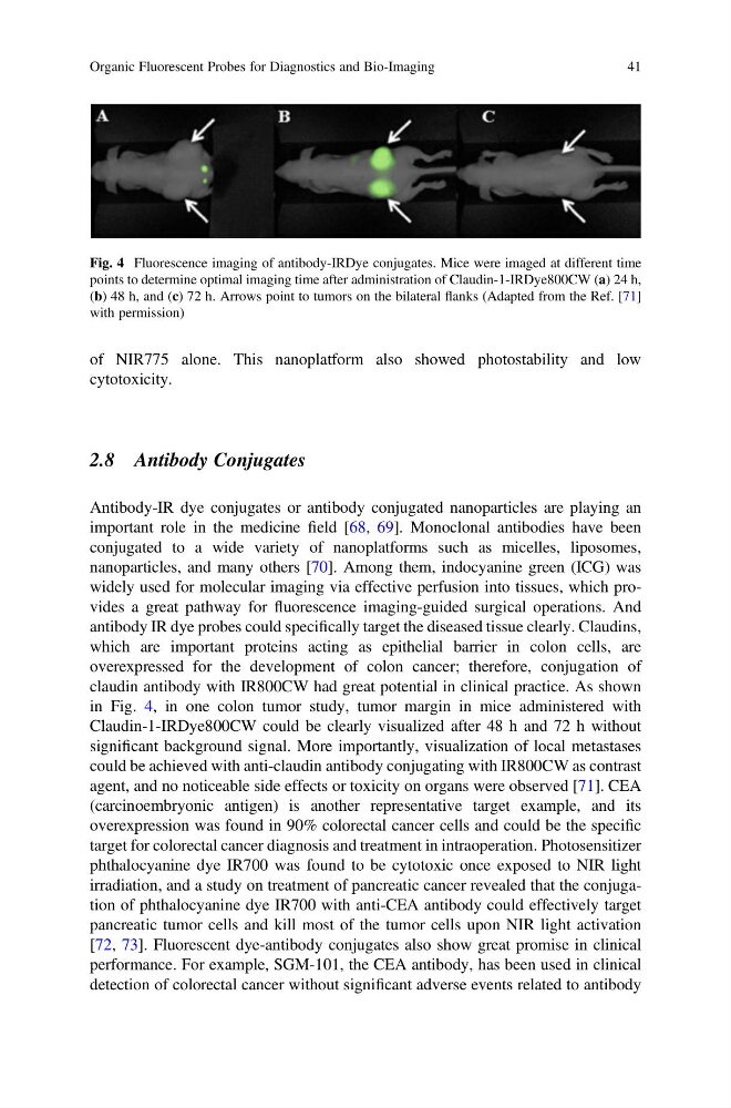
claudin antibody with IR800CW had great potential in clinical practice. As shown
in Fig. 4, in one colon tumor study, tumor margin in mice administered with
Claudin-1-IRDye800CW could be clearly visualized after 48 h and 72 h without
significant background signal. More importantly, visualization of local metastases
could be achieved with anti-claudin antibody conjugating with IR800CW as contrast
agent, and no noticeable side effects or toxicity on organs were observed [71]. CEA
(carcinoembryonic antigen) is another representative target example, and its
overexpression was found in 90% colorectal cancer cells and could be the specific
target for colorectal cancer diagnosis and treatment in intraoperation. Photosensitizer
phthalocyanine dye IR700 was found to be cytotoxic once exposed to NIR light
irradiation, and a study on treatment of pancreatic cancer revealed that the conjugation of phthalocyanine dye IR700 with anti-CEA antibody could effectively target
pancreatic tumor cells and kill most of the tumor cells upon NIR light activation
[72, 73]. Fluorescent dye-antibody conjugates also show great promise in clinical
performance. For example, SGM-101, the CEA antibody, has been used in clinical
detection of colorectal cancer without significant adverse events related to antibody
Fig. 4 Fluorescence imaging of antibody-IRDye conjugates. Mice were imaged at different time
points to determine optimal imaging time after administration of Claudin-1-IRDye800CW (a) 24 h,
(b) 48 h, and (c) 72 h. Arrows point to tumors on the bilateral flanks (Adapted from the Ref. [71]
with permission)
Organic Fluorescent Probes for Diagnostics and Bio-Imaging
41
cytotoxicity.
2.8 Antibody Conjugates
Antibody-IR dye conjugates or antibody conjugated nanoparticles are playing an
important role in the medicine field [68, 69]. Monoclonal antibodies have been
conjugated to a wide variety of nanoplatforms such as micelles, liposomes,
nanoparticles, and many others [70]. Among them, indocyanine green (ICG) was
widely used for molecular imaging via effective perfusion into tissues, which provides a great pathway for fluorescence imaging-guided surgical operations. And
antibody IR dye probes could specifically target the diseased tissue clearly. Claudins,
which are important proteins acting as epithelial barrier in colon cells, are
overexpressed for the development of colon cancer; therefore, conjugation of
claudin antibody with IR800CW had great potential in clinical practice. As shown
in Fig. 4, in one colon tumor study, tumor margin in mice administered with
Claudin-1-IRDye800CW could be clearly visualized after 48 h and 72 h without
significant background signal. More importantly, visualization of local metastases
could be achieved with anti-claudin antibody conjugating with IR800CW as contrast
agent, and no noticeable side effects or toxicity on organs were observed [71]. CEA
(carcinoembryonic antigen) is another representative target example, and its
overexpression was found in 90% colorectal cancer cells and could be the specific
target for colorectal cancer diagnosis and treatment in intraoperation. Photosensitizer
phthalocyanine dye IR700 was found to be cytotoxic once exposed to NIR light
irradiation, and a study on treatment of pancreatic cancer revealed that the conjugation of phthalocyanine dye IR700 with anti-CEA antibody could effectively target
pancreatic tumor cells and kill most of the tumor cells upon NIR light activation
[72, 73]. Fluorescent dye-antibody conjugates also show great promise in clinical
performance. For example, SGM-101, the CEA antibody, has been used in clinical
detection of colorectal cancer without significant adverse events related to antibody
Fig. 4 Fluorescence imaging of antibody-IRDye conjugates. Mice were imaged at different time
points to determine optimal imaging time after administration of Claudin-1-IRDye800CW (a) 24 h,
(b) 48 h, and (c) 72 h. Arrows point to tumors on the bilateral flanks (Adapted from the Ref. [71]
with permission)
Organic Fluorescent Probes for Diagnostics and Bio-Imaging
41