

shown improvement in detection of depressed-type gastric neoplastic lesions [9] but
not in detection of adenomatous colon polyps [10].
Fluorescence Imaging Modalities Fluorescence imaging is an emerging and
promising avenue for the present and future of clinical imaging, including in
endoscopic applications. Analogous to WLE in visible-spectrum light imaging, the
most widely used form of fluorescence imaging in endoscopic procedures is
autofluorescence endoscopy (AFE). AFE endoscopes exploit differences in the
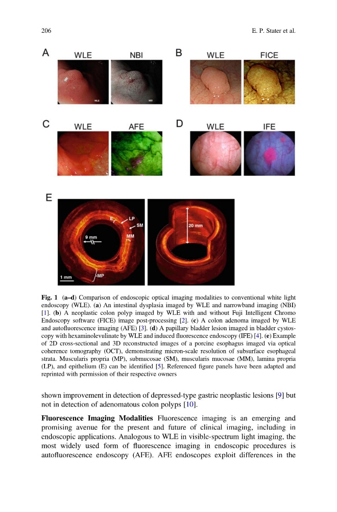
Fig. 1 (a–d) Comparison of endoscopic optical imaging modalities to conventional white light
endoscopy (WLE). (a) An intestinal dysplasia imaged by WLE and narrowband imaging (NBI)
[1]. (b) A neoplastic colon polyp imaged by WLE with and without Fuji Intelligent Chromo
Endoscopy software (FICE) image post-processing [2]. (c) A colon adenoma imaged by WLE
and autofluorescence imaging (AFE) [3]. (d) A papillary bladder lesion imaged in bladder cystoscopy with hexaminolevulinate by WLE and induced fluorescence endoscopy (IFE) [4]. (e) Example
of 2D cross-sectional and 3D reconstructed images of a porcine esophagus imaged via optical
coherence tomography (OCT), demonstrating micron-scale resolution of subsurface esophageal
strata. Muscularis propria (MP), submucosae (SM), muscularis mucosae (MM), lamina propria
(LP), and epithelium (E) can be identified [5]. Referenced figure panels have been adapted and
reprinted with permission of their respective owners
206
E. P. Stater et al.
not in detection of adenomatous colon polyps [10].
Fluorescence Imaging Modalities Fluorescence imaging is an emerging and
promising avenue for the present and future of clinical imaging, including in
endoscopic applications. Analogous to WLE in visible-spectrum light imaging, the
most widely used form of fluorescence imaging in endoscopic procedures is
autofluorescence endoscopy (AFE). AFE endoscopes exploit differences in the
Fig. 1 (a–d) Comparison of endoscopic optical imaging modalities to conventional white light
endoscopy (WLE). (a) An intestinal dysplasia imaged by WLE and narrowband imaging (NBI)
[1]. (b) A neoplastic colon polyp imaged by WLE with and without Fuji Intelligent Chromo
Endoscopy software (FICE) image post-processing [2]. (c) A colon adenoma imaged by WLE
and autofluorescence imaging (AFE) [3]. (d) A papillary bladder lesion imaged in bladder cystoscopy with hexaminolevulinate by WLE and induced fluorescence endoscopy (IFE) [4]. (e) Example
of 2D cross-sectional and 3D reconstructed images of a porcine esophagus imaged via optical
coherence tomography (OCT), demonstrating micron-scale resolution of subsurface esophageal
strata. Muscularis propria (MP), submucosae (SM), muscularis mucosae (MM), lamina propria
(LP), and epithelium (E) can be identified [5]. Referenced figure panels have been adapted and
reprinted with permission of their respective owners
206
E. P. Stater et al.