

activity in vitro and in vivo is vitally required for cancer diagnosis. Moreover, at the
side of the single-wavelength fluorescence-intensity-based systems, ratiometric
fluorescence probes are of crucial practical advantages like enhanced signal-tobackground ratio, in which the detectable ratio signal can be obtained via two
isolated read-out channels of activated versus unreacted probes and bringing about
improved and reliable signal quantification.
Nanomaterials has been regarded as one of most emerging biomedical imaging
probes because of their special properties are being more and more studied for the
detection of β-gal. Compared to conventional fluorescent probes, nanomaterials
possess unique superiority such as smaller size, higher selectivity, and better biocompatibility. For example, pH-driven-luminescence GSH-protected CuNCs with
AIE property and long decay time are promising to monitor β-gal continuously, and
GOS-capped MSNs loaded with certain drugs are potential probes targeting SA-β-gal
as well as device for drug delivery. Another used nanoprobe is β-CD-CQDs, which is
based on a combined host-guest recognition and specific static quenching-induced
signal transduction mechanism, because of their stable fluorescence emitting, excellent
photostability, and low detection limit.
There are a number of fluorogenic probes being developed for detecting β-gal, but
their applications are limited by a few drawbacks such as poor cellular permeability,
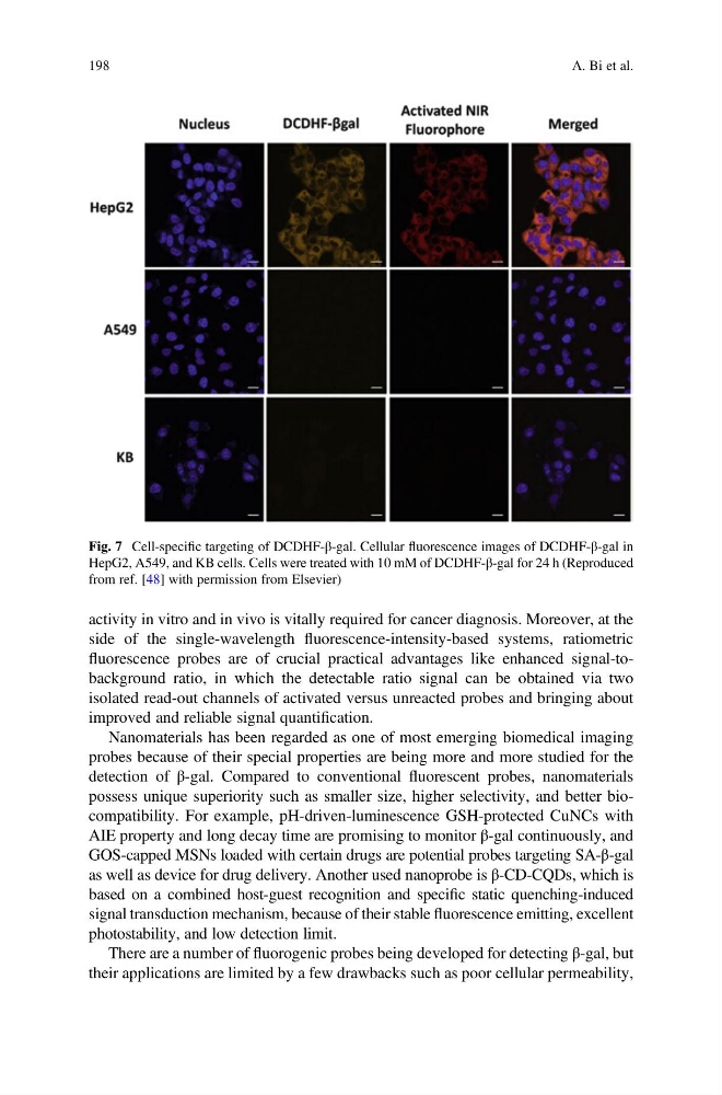
Fig. 7 Cell-specific targeting of DCDHF-β-gal. Cellular fluorescence images of DCDHF-β-gal in
HepG2, A549, and KB cells. Cells were treated with 10 mM of DCDHF-β-gal for 24 h (Reproduced
from ref. [48] with permission from Elsevier)
198
A. Bi et al.
side of the single-wavelength fluorescence-intensity-based systems, ratiometric
fluorescence probes are of crucial practical advantages like enhanced signal-tobackground ratio, in which the detectable ratio signal can be obtained via two
isolated read-out channels of activated versus unreacted probes and bringing about
improved and reliable signal quantification.
Nanomaterials has been regarded as one of most emerging biomedical imaging
probes because of their special properties are being more and more studied for the
detection of β-gal. Compared to conventional fluorescent probes, nanomaterials
possess unique superiority such as smaller size, higher selectivity, and better biocompatibility. For example, pH-driven-luminescence GSH-protected CuNCs with
AIE property and long decay time are promising to monitor β-gal continuously, and
GOS-capped MSNs loaded with certain drugs are potential probes targeting SA-β-gal
as well as device for drug delivery. Another used nanoprobe is β-CD-CQDs, which is
based on a combined host-guest recognition and specific static quenching-induced
signal transduction mechanism, because of their stable fluorescence emitting, excellent
photostability, and low detection limit.
There are a number of fluorogenic probes being developed for detecting β-gal, but
their applications are limited by a few drawbacks such as poor cellular permeability,
Fig. 7 Cell-specific targeting of DCDHF-β-gal. Cellular fluorescence images of DCDHF-β-gal in
HepG2, A549, and KB cells. Cells were treated with 10 mM of DCDHF-β-gal for 24 h (Reproduced
from ref. [48] with permission from Elsevier)
198
A. Bi et al.