

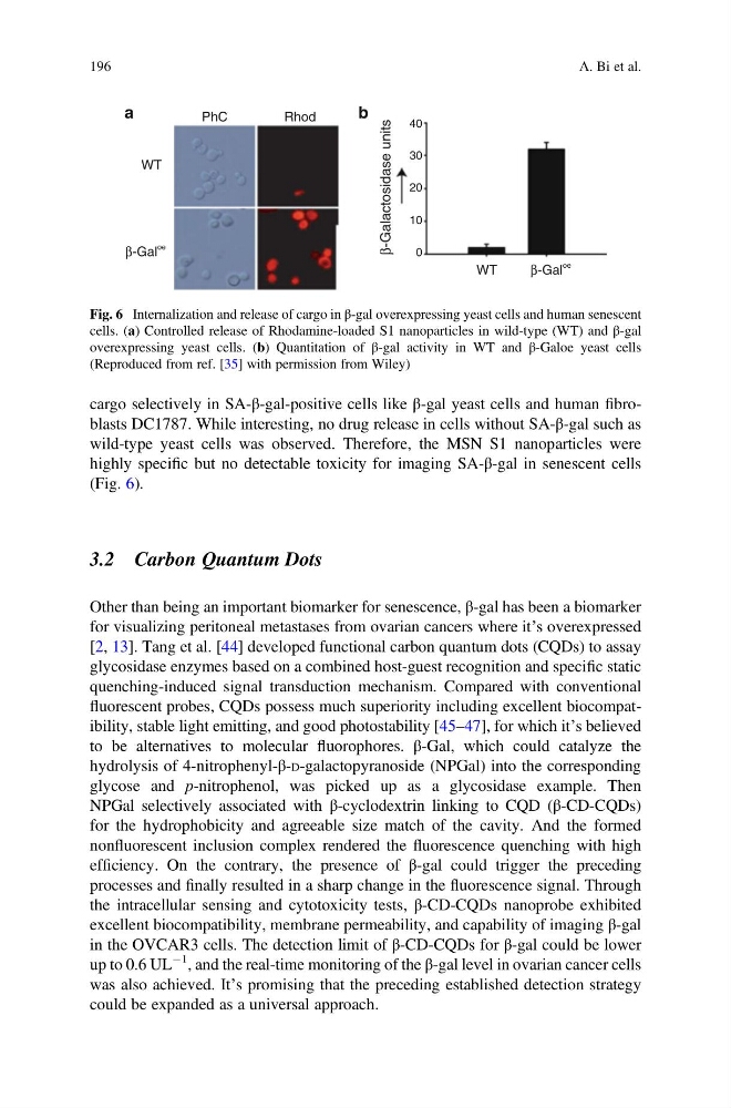
cargo selectively in SA-β-gal-positive cells like β-gal yeast cells and human fibroblasts DC1787. While interesting, no drug release in cells without SA-β-gal such as
wild-type yeast cells was observed. Therefore, the MSN S1 nanoparticles were
highly specific but no detectable toxicity for imaging SA-β-gal in senescent cells
(Fig. 6).
3.2 Carbon Quantum Dots
Other than being an important biomarker for senescence, β-gal has been a biomarker
for visualizing peritoneal metastases from ovarian cancers where it’s overexpressed
[2, 13]. Tang et al. [44] developed functional carbon quantum dots (CQDs) to assay
glycosidase enzymes based on a combined host-guest recognition and specific static
quenching-induced signal transduction mechanism. Compared with conventional
fluorescent probes, CQDs possess much superiority including excellent biocompatibility, stable light emitting, and good photostability [45–47], for which it’s believed
to be alternatives to molecular fluorophores. β-Gal, which could catalyze the
hydrolysis of 4-nitrophenyl-β-D-galactopyranoside (NPGal) into the corresponding
glycose and p-nitrophenol, was picked up as a glycosidase example. Then
NPGal selectively associated with β-cyclodextrin linking to CQD (β-CD-CQDs)
for the hydrophobicity and agreeable size match of the cavity. And the formed
nonfluorescent inclusion complex rendered the fluorescence quenching with high
efficiency. On the contrary, the presence of β-gal could trigger the preceding
processes and finally resulted in a sharp change in the fluorescence signal. Through
the intracellular sensing and cytotoxicity tests, β-CD-CQDs nanoprobe exhibited
excellent biocompatibility, membrane permeability, and capability of imaging β-gal
in the OVCAR3 cells. The detection limit of β-CD-CQDs for β-gal could be lower
up to 0.6 UL
À1 , and the real-time monitoring of the β-gal level in ovarian cancer cells
was also achieved. It’s promising that the preceding established detection strategy
could be expanded as a universal approach.
a
b
PhC
Rhod
40
30
20
10
0
β-Gal
oe
β-Gal
oe
β-Galactosidase units
WT
WT
Fig. 6 Internalization and release of cargo in β-gal overexpressing yeast cells and human senescent
cells. (a) Controlled release of Rhodamine-loaded S1 nanoparticles in wild-type (WT) and β-gal
overexpressing yeast cells. (b) Quantitation of β-gal activity in WT and β-Galoe yeast cells
(Reproduced from ref. [35] with permission from Wiley)
196
A. Bi et al.
wild-type yeast cells was observed. Therefore, the MSN S1 nanoparticles were
highly specific but no detectable toxicity for imaging SA-β-gal in senescent cells
(Fig. 6).
3.2 Carbon Quantum Dots
Other than being an important biomarker for senescence, β-gal has been a biomarker
for visualizing peritoneal metastases from ovarian cancers where it’s overexpressed
[2, 13]. Tang et al. [44] developed functional carbon quantum dots (CQDs) to assay
glycosidase enzymes based on a combined host-guest recognition and specific static
quenching-induced signal transduction mechanism. Compared with conventional
fluorescent probes, CQDs possess much superiority including excellent biocompatibility, stable light emitting, and good photostability [45–47], for which it’s believed
to be alternatives to molecular fluorophores. β-Gal, which could catalyze the
hydrolysis of 4-nitrophenyl-β-D-galactopyranoside (NPGal) into the corresponding
glycose and p-nitrophenol, was picked up as a glycosidase example. Then
NPGal selectively associated with β-cyclodextrin linking to CQD (β-CD-CQDs)
for the hydrophobicity and agreeable size match of the cavity. And the formed
nonfluorescent inclusion complex rendered the fluorescence quenching with high
efficiency. On the contrary, the presence of β-gal could trigger the preceding
processes and finally resulted in a sharp change in the fluorescence signal. Through
the intracellular sensing and cytotoxicity tests, β-CD-CQDs nanoprobe exhibited
excellent biocompatibility, membrane permeability, and capability of imaging β-gal
in the OVCAR3 cells. The detection limit of β-CD-CQDs for β-gal could be lower
up to 0.6 UL
À1 , and the real-time monitoring of the β-gal level in ovarian cancer cells
was also achieved. It’s promising that the preceding established detection strategy
could be expanded as a universal approach.
a
b
PhC
Rhod
40
30
20
10
0
β-Gal
oe
β-Gal
oe
β-Galactosidase units
WT
WT
Fig. 6 Internalization and release of cargo in β-gal overexpressing yeast cells and human senescent
cells. (a) Controlled release of Rhodamine-loaded S1 nanoparticles in wild-type (WT) and β-gal
overexpressing yeast cells. (b) Quantitation of β-gal activity in WT and β-Galoe yeast cells
(Reproduced from ref. [35] with permission from Wiley)
196
A. Bi et al.