

exposing to β-gal, the carbamate compound was cleaved to afford the amino group
as the electron donor. An internal charge transfer (ICT) effect was observed to shift
the emission to the stronger fluorescent emission. After treating with β-gal in PBS
buffer (10 mM, pH 7.4, 37
C), the fluorescent intensity of a 1 μM probe solution
increased gradually at 540 nm while decreased at 460 nm. The ratio of the emission
intensities at 410–460 nm and 520–570 nm increased by 120-fold upon reaction.
Meanwhile, D-galactose, a well-known competitive inhibitor of β-gal, could not
trigger this fluorescent emission, indicating that the fluorescent enhancement is
specific to β-gal activity. A linear relationship was observed against the β-gal
concentration ranging from 0 to 2.0 nM, indicating that probe 1 could quantify
β-gal in the low concentration range. Next, the application practicability of 1 has
been examined in a representative cell senescence model via monitoring the endogenous SA-β-gal activity. Doubling times and population were recorded in this
process, during the middle stages of senescence, suggesting that the probe could
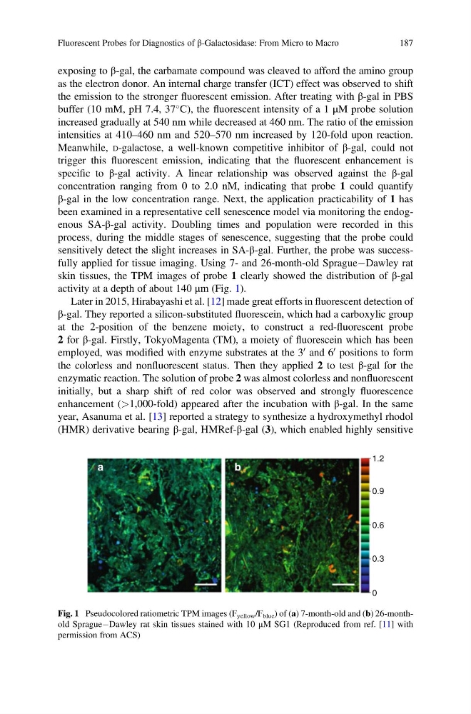
sensitively detect the slight increases in SA-β-gal. Further, the probe was successfully applied for tissue imaging. Using 7- and 26-month-old SpragueÀDawley rat
skin tissues, the TPM images of probe 1 clearly showed the distribution of β-gal
activity at a depth of about 140 μm (Fig. 1).
Later in 2015, Hirabayashi et al. [12] made great efforts in fluorescent detection of
β-gal. They reported a silicon-substituted fluorescein, which had a carboxylic group
at the 2-position of the benzene moiety, to construct a red-fluorescent probe
2 for β-gal. Firstly, TokyoMagenta (TM), a moiety of fluorescein which has been
employed, was modified with enzyme substrates at the 3
0 and 6
0 positions to form
the colorless and nonfluorescent status. Then they applied 2 to test β-gal for the
enzymatic reaction. The solution of probe 2 was almost colorless and nonfluorescent
initially, but a sharp shift of red color was observed and strongly fluorescence
enhancement (>1,000-fold) appeared after the incubation with β-gal. In the same
year, Asanuma et al. [13] reported a strategy to synthesize a hydroxymethyl rhodol
(HMR) derivative bearing β-gal, HMRef-β-gal (3), which enabled highly sensitive
a
b
0.9
1.2
0.6
0.3
0
Fig. 1 Pseudocolored ratiometric TPM images (F yellow /F blue ) of (a) 7-month-old and (b) 26-monthold SpragueÀDawley rat skin tissues stained with 10 μM SG1 (Reproduced from ref. [11] with
permission from ACS)
Fluorescent Probes for Diagnostics of β-Galactosidase: From Micro to Macro
187
as the electron donor. An internal charge transfer (ICT) effect was observed to shift
the emission to the stronger fluorescent emission. After treating with β-gal in PBS
buffer (10 mM, pH 7.4, 37
C), the fluorescent intensity of a 1 μM probe solution
increased gradually at 540 nm while decreased at 460 nm. The ratio of the emission
intensities at 410–460 nm and 520–570 nm increased by 120-fold upon reaction.
Meanwhile, D-galactose, a well-known competitive inhibitor of β-gal, could not
trigger this fluorescent emission, indicating that the fluorescent enhancement is
specific to β-gal activity. A linear relationship was observed against the β-gal
concentration ranging from 0 to 2.0 nM, indicating that probe 1 could quantify
β-gal in the low concentration range. Next, the application practicability of 1 has
been examined in a representative cell senescence model via monitoring the endogenous SA-β-gal activity. Doubling times and population were recorded in this
process, during the middle stages of senescence, suggesting that the probe could
sensitively detect the slight increases in SA-β-gal. Further, the probe was successfully applied for tissue imaging. Using 7- and 26-month-old SpragueÀDawley rat
skin tissues, the TPM images of probe 1 clearly showed the distribution of β-gal
activity at a depth of about 140 μm (Fig. 1).
Later in 2015, Hirabayashi et al. [12] made great efforts in fluorescent detection of
β-gal. They reported a silicon-substituted fluorescein, which had a carboxylic group
at the 2-position of the benzene moiety, to construct a red-fluorescent probe
2 for β-gal. Firstly, TokyoMagenta (TM), a moiety of fluorescein which has been
employed, was modified with enzyme substrates at the 3
0 and 6
0 positions to form
the colorless and nonfluorescent status. Then they applied 2 to test β-gal for the
enzymatic reaction. The solution of probe 2 was almost colorless and nonfluorescent
initially, but a sharp shift of red color was observed and strongly fluorescence
enhancement (>1,000-fold) appeared after the incubation with β-gal. In the same
year, Asanuma et al. [13] reported a strategy to synthesize a hydroxymethyl rhodol
(HMR) derivative bearing β-gal, HMRef-β-gal (3), which enabled highly sensitive
a
b
0.9
1.2
0.6
0.3
0
Fig. 1 Pseudocolored ratiometric TPM images (F yellow /F blue ) of (a) 7-month-old and (b) 26-monthold SpragueÀDawley rat skin tissues stained with 10 μM SG1 (Reproduced from ref. [11] with
permission from ACS)
Fluorescent Probes for Diagnostics of β-Galactosidase: From Micro to Macro
187